Antibody-Dependent Enhanced (ADE) Immunity

The method to the dengue virus's maddening infectiousness.

Written byBeth Marie Mole
| 1 min read

Register for free to listen to this article
Listen with Speechify
0:00
1:00
Share

View full size JPG | PDFTAMI TOLPA

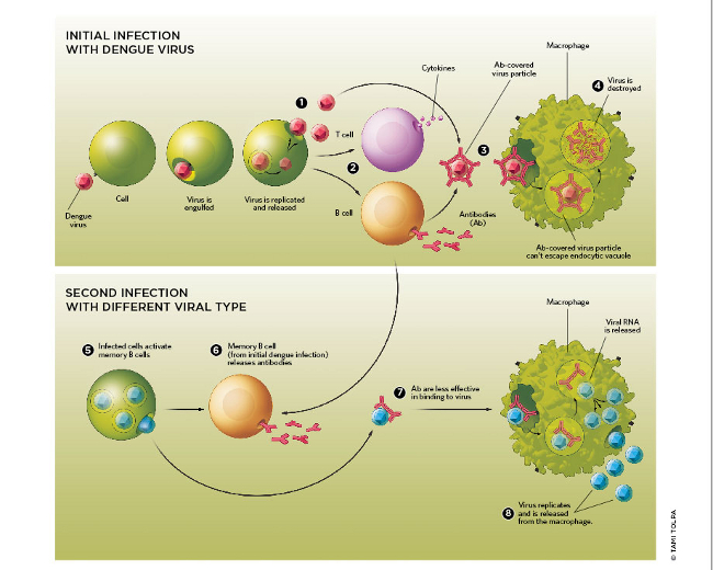
Dengue infects by attaching to a cell that then engulfs the virus in a vacuole via endocytosis. The virus rearranges its coat proteins to bind to the vacuole membrane, releasing its capsid and genome into the cytoplasm, where it is replicated and packaged into newly made virions (1). The infected cell also triggers an immune reaction (2) that includes the recruitment of T cells that release pro-inflammatory cytokines and B cells that generate antibodies. These antibodies are specific for the infecting virus, and bind and cover dengue’s coat (3). When this happens, macrophages and monocytes clear the virus from the bloodstream by binding to and engulfing the antibody-coated viruses—which can no longer escape the endocytic vacuole—and destroying them (4).

When a person is infected with ...

Interested in reading more?

Become a Member of

The Scientist Logo
Receive full access to more than 35 years of archives, as well as TS Digest, digital editions of The Scientist, feature stories, and much more!
Already a member? Login Here
Illustration of a developing fetus surrounded by a clear fluid with a subtle yellow tinge, representing amniotic fluid.
January 2026, Issue 1

What Is the Amniotic Fluid Composed of?

The liquid world of fetal development provides a rich source of nutrition and protection tailored to meet the needs of the growing fetus.

View this Issue
Skip the Wait for Protein Stability Data with Aunty

Skip the Wait for Protein Stability Data with Aunty

Unchained Labs
Graphic of three DNA helices in various colors

An Automated DNA-to-Data Framework for Production-Scale Sequencing

illumina
Exploring Cellular Organization with Spatial Proteomics

Exploring Cellular Organization with Spatial Proteomics

Abstract illustration of spheres with multiple layers, representing endoderm, ectoderm, and mesoderm derived organoids

Organoid Origins and How to Grow Them

Thermo Fisher Logo

Products

nuclera logo

Nuclera eProtein Discovery System installed at leading Universities in Taiwan

Brandtech Logo

BRANDTECH Scientific Introduces the Transferpette® pro Micropipette: A New Twist on Comfort and Control

Biotium Logo

Biotium Launches GlycoLiner™ Cell Surface Glycoprotein Labeling Kits for Rapid and Selective Cell Surface Imaging

Colorful abstract spiral dot pattern on a black background

Thermo Scientific X and S Series General Purpose Centrifuges

Thermo Fisher Logo