Chromatin Conformation Computed

By manipulating DNA sequences that guide genome-folding, researchers confirm an existing model of chromatin structure inside the nucleus.

Written byAnna Azvolinsky
| 4 min read

Register for free to listen to this article
Listen with Speechify
0:00
4:00
Share

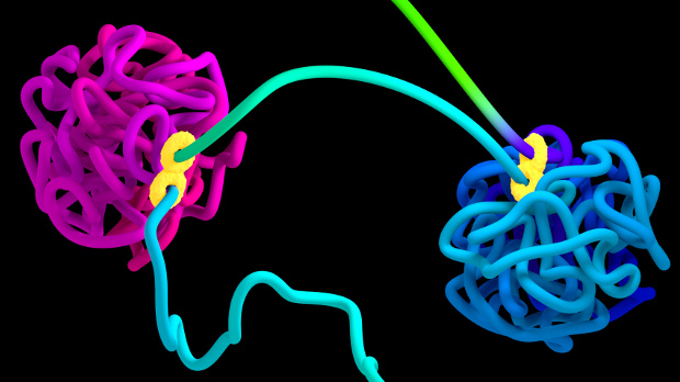
Two chromatin loops (magenta and blue) held together by a CTCF-containing protein complex (yellow)ADRIAN SANBORN, NAJEEB TARAZI, EREZ LIEBERMAN AIDEN, BAYLOR COLLEGE OF MEDICINE

Compared to its sequence, relatively little is known about the structure of the human genome, which enables more than two meters of chromatin to fit inside the nucleus. Chromosomes are thought to be organized into loops that bring together distant DNA elements—genes, promoters, and enhancers. These chromatin loops are thought to help facilitate gene regulation.

Now, researchers at the Baylor College of Medicine’s Center for Genome Architecture in Houston, Texas, have developed a mathematical model using the binding pattern of a single protein to DNA to predict the looping organization of the human genome. The model helped the team predict the results of its CRISPR-based experimental modification of some of these protein-binding sites in human cells. The team’s results, published this week (October 19) in ...

Interested in reading more?

Become a Member of

The Scientist Logo
Receive full access to more than 35 years of archives, as well as TS Digest, digital editions of The Scientist, feature stories, and much more!
Already a member? Login Here

Related Topics

Meet the Author

  • head shot of blond woman wearing glasses

    Anna Azvolinsky received a PhD in molecular biology in November 2008 from Princeton University. Her graduate research focused on a genome-wide analyses of genomic integrity and DNA replication. She did a one-year post-doctoral fellowship at Memorial Sloan Kettering Cancer Center in New York City and then left academia to pursue science writing. She has been a freelance science writer since 2012, based in New York City.

    View Full Profile
Share
Illustration of a developing fetus surrounded by a clear fluid with a subtle yellow tinge, representing amniotic fluid.
January 2026, Issue 1

What Is the Amniotic Fluid Composed of?

The liquid world of fetal development provides a rich source of nutrition and protection tailored to meet the needs of the growing fetus.

View this Issue
Skip the Wait for Protein Stability Data with Aunty

Skip the Wait for Protein Stability Data with Aunty

Unchained Labs
Graphic of three DNA helices in various colors

An Automated DNA-to-Data Framework for Production-Scale Sequencing

illumina
Exploring Cellular Organization with Spatial Proteomics

Exploring Cellular Organization with Spatial Proteomics

Abstract illustration of spheres with multiple layers, representing endoderm, ectoderm, and mesoderm derived organoids

Organoid Origins and How to Grow Them

Thermo Fisher Logo

Products

Brandtech Logo

BRANDTECH Scientific Introduces the Transferpette® pro Micropipette: A New Twist on Comfort and Control

Biotium Logo

Biotium Launches GlycoLiner™ Cell Surface Glycoprotein Labeling Kits for Rapid and Selective Cell Surface Imaging

Colorful abstract spiral dot pattern on a black background

Thermo Scientific X and S Series General Purpose Centrifuges

Thermo Fisher Logo
Abstract background with red and blue laser lights

VANTAstar Flexible microplate reader with simplified workflows

BMG LABTECH