Combating Coronaviruses

Scientists discover a molecule that defeats MERS and other coronaviruses in human cells.

Written byRuth Williams
| 3 min read

Register for free to listen to this article
Listen with Speechify
0:00
3:00
Share

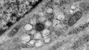
Coronavirus replication vesicles inside host cellVOLKER THIEL, EDWARD TRYBALA Coronaviruses, including the one responsible for the ongoing outbreak of Middle East respiratory syndrome (MERS), can be defeated in a dish by a molecule that destroys their replication vesicles, according to a paper published in PLOS Pathogens today (May 29). The molecule, called K22, is the first antiviral substance known to interfere with such vesicles, revealing them as a novel exploitable weak point in the coronavirus (CoV) life cycle.

“There’s no way to treat a virus with just one drug. You need to come at it from a couple of different angles, and this is a brand new fresh angle from which to attack a dangerous pathogen,” said Benjamin Neuman, a molecular virologist at the University of Reading in the U.K., who was not involved in the study. “And it seems to work on all the coronaviruses they tested, which is lovely.”

Until a decade ago, CoVs were not considered a significant threat to human health because the few that did infect humans caused non-life-threatening upper respiratory tract infections (essentially, colds). But then, in 2003, the SARS CoV emerged and killed nearly 10 percent of the people it infected. And the MERS-CoV that’s currently ...

Interested in reading more?

Become a Member of

The Scientist Logo
Receive full access to more than 35 years of archives, as well as TS Digest, digital editions of The Scientist, feature stories, and much more!
Already a member? Login Here

Related Topics

Meet the Author

  • ruth williams

    Ruth is a freelance journalist. Before freelancing, Ruth was a news editor for the Journal of Cell Biology in New York and an assistant editor for Nature Reviews Neuroscience in London. Prior to that, she was a bona fide pipette-wielding, test tube–shaking, lab coat–shirking research scientist. She has a PhD in genetics from King’s College London, and was a postdoc in stem cell biology at Imperial College London. Today she lives and writes in Connecticut.

    View Full Profile
Share
Illustration of a developing fetus surrounded by a clear fluid with a subtle yellow tinge, representing amniotic fluid.
January 2026, Issue 1

What Is the Amniotic Fluid Composed of?

The liquid world of fetal development provides a rich source of nutrition and protection tailored to meet the needs of the growing fetus.

View this Issue
Skip the Wait for Protein Stability Data with Aunty

Skip the Wait for Protein Stability Data with Aunty

Unchained Labs
Graphic of three DNA helices in various colors

An Automated DNA-to-Data Framework for Production-Scale Sequencing

illumina
Exploring Cellular Organization with Spatial Proteomics

Exploring Cellular Organization with Spatial Proteomics

Abstract illustration of spheres with multiple layers, representing endoderm, ectoderm, and mesoderm derived organoids

Organoid Origins and How to Grow Them

Thermo Fisher Logo

Products

Brandtech Logo

BRANDTECH Scientific Introduces the Transferpette® pro Micropipette: A New Twist on Comfort and Control

Biotium Logo

Biotium Launches GlycoLiner™ Cell Surface Glycoprotein Labeling Kits for Rapid and Selective Cell Surface Imaging

Colorful abstract spiral dot pattern on a black background

Thermo Scientific X and S Series General Purpose Centrifuges

Thermo Fisher Logo
Abstract background with red and blue laser lights

VANTAstar Flexible microplate reader with simplified workflows

BMG LABTECH