
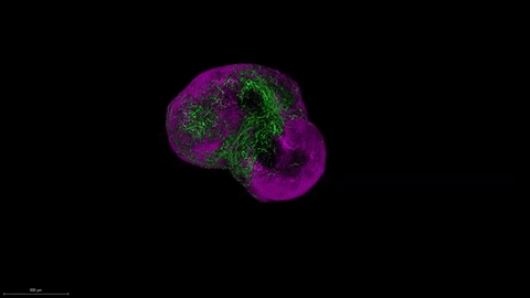
 Fused human forebrain spheroids, iDISCO 3-D reconstructionPASCA LABScientists know little about the early development of the human brain. As a result, they also have limited data about how human brain development might relate to neuropsychiatric disorders, such as autism and schizophrenia. Since 2013, however, scientists having been studying the developing human brain using neurons derived from human induced pluripotent stem cells (iPSCs), which are cultured in three dimensions into pea-size structures that mimic the full organ. Two studies published today (April 26) in Nature advance these research methods. In one paper, Harvard’s Paula Arlotta and colleagues described the development of organoids, or “mini brains.” In the other, Stanford’s Sergiu Pasca and colleagues used neural spheroids—balls of tissue containing more than a million neurons each—to study the interactions of two brain regions crucial to the development of the cerebral cortex.
“The major conclusion is the confirmation/validation that the human pluripotent stem cells are plastic enough to generate the diversity of cells necessary to recreate human, early stages of neurodevelopment in a dish,” Alysson Muotri, who studies neurological diseases using iPSCs at University of California, San Diego, but was not involved in either study, told The Scientist in an email. “Every neuroscientist working with early brain development will be excited by reading these articles.”
Researchers hope to use brain organoids and spheroids to study neurodevelopment and neuropsychiatric disorders; these mini brains have already been used to study Zika virus infection–linked microcephaly and autism spectrum disorders.
Because many neuropsychiatric disorders are influenced by a person’s genetics, it is difficult to study these diseases in standard animal models. ...


