Enhancers Drive Pancreatic Cancer Metastasis: Study

Mouse organoids reveal that a protein active during embryonic development joins forces with gene enhancers to revert cancer cells to an earlier developmental state.

Written byDiana Kwon
| 2 min read

Register for free to listen to this article
Listen with Speechify
0:00
2:00
Share

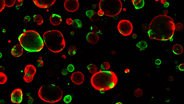
Organoids grown from pancreatic tissue. Red-colored organoids are composed of normal cells; green organoids are grown from pancreatic tumor samples.TUVESON LAB, CSHLPancreatic ductal adenocarcinoma (PDA), a common form of pancreatic cancer, is often fatal due to its high tendency to metastasize. In a study published today (July 27) in Cell, scientists have uncovered a mechanism in mice that may be driving the spread of this cancer.

To examine the changes that promote cancer metastasis, researchers first developed organoids using primary tumor and metastatic cells from a mouse model of PDA. By comparing these mini organs, the team found that metastatic organoids had more active enhancers, short DNA sequences that bind to transcription factors to enhance gene expression, than the ones derived from primary tumor cells.

Further analysis revealed that FOXA1, a protein active during embryonic development, was binding to these enhancers. In addition, cells with higher levels of this molecule displayed an increased expression of genes found during earlier developmental states.

“We show that to metastasize, the cell has to change, in effect, its whole telecommunications network—its enhancers are being reprogrammed,” study co-author Christopher Vakoc, a cancer epigenetics researchers at Cold ...

Interested in reading more?

Become a Member of

The Scientist Logo
Receive full access to more than 35 years of archives, as well as TS Digest, digital editions of The Scientist, feature stories, and much more!
Already a member? Login Here

Related Topics

Meet the Author

  • Diana is a freelance science journalist who covers the life sciences, health, and academic life. She’s a regular contributor to The Scientist and her work has appeared in several other publications, including Scientific American, Knowable, and Quanta. Diana was a former intern at The Scientist and she holds a master’s degree in neuroscience from McGill University. She’s currently based in Berlin, Germany.

    View Full Profile
Share
Illustration of a developing fetus surrounded by a clear fluid with a subtle yellow tinge, representing amniotic fluid.
January 2026, Issue 1

What Is the Amniotic Fluid Composed of?

The liquid world of fetal development provides a rich source of nutrition and protection tailored to meet the needs of the growing fetus.

View this Issue
Skip the Wait for Protein Stability Data with Aunty

Skip the Wait for Protein Stability Data with Aunty

Unchained Labs
Graphic of three DNA helices in various colors

An Automated DNA-to-Data Framework for Production-Scale Sequencing

illumina
Exploring Cellular Organization with Spatial Proteomics

Exploring Cellular Organization with Spatial Proteomics

Abstract illustration of spheres with multiple layers, representing endoderm, ectoderm, and mesoderm derived organoids

Organoid Origins and How to Grow Them

Thermo Fisher Logo

Products

Brandtech Logo

BRANDTECH Scientific Introduces the Transferpette® pro Micropipette: A New Twist on Comfort and Control

Biotium Logo

Biotium Launches GlycoLiner™ Cell Surface Glycoprotein Labeling Kits for Rapid and Selective Cell Surface Imaging

Colorful abstract spiral dot pattern on a black background

Thermo Scientific X and S Series General Purpose Centrifuges

Thermo Fisher Logo
Abstract background with red and blue laser lights

VANTAstar Flexible microplate reader with simplified workflows

BMG LABTECH