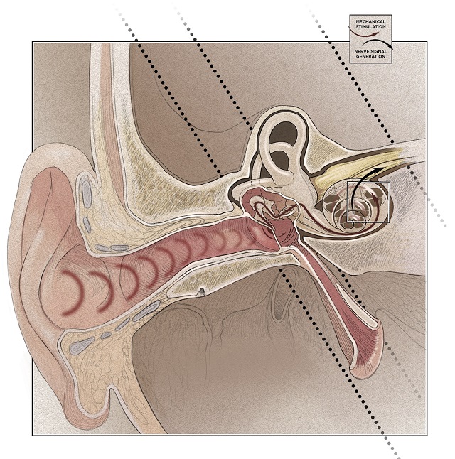
When sound enters the ear canal, it vibrates the tympanic membrane, or eardrum. These vibrations are passed through the inner ear via three small bones called ossicles: the malleus, the incus, and the stapes. Finally, vibrations of the stapes stimulate the movement of a fluid called perilymph within the bony labyrinth of the inner ear.
 See labeled infographic: JPG© CATHERINE DELPHIA
Perilymph fills the both the vestibular and tympanic ducts of the cochlea. Between these two channels lies the cochlear duct, which is home to the organ of Corti. There, the soundinduced movement of perilymph in the cochlea is translated to an electrical signal that is sent to the brain for processing.
An electrical signal is generated by inner hair cells that sit above the basilar membrane, which separates the cochlear ...


