Monitoring Mitochondrial Mutations

Induced pluripotent stem cells—particularly those generated from older patients—should be screened for defects in mitochondrial DNA, a study shows.

Written byCatherine Offord
| 2 min read

Register for free to listen to this article
Listen with Speechify
0:00
2:00
Share

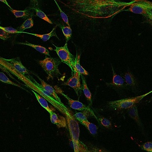
Human fibroblasts derived from embryonic stem cells showing nuclei (blue) and mitochondria (red)SHOUKHRAT MITALIPOVThe number of mutations in mitochondrial DNA (mtDNA) could vary substantially between different lines of induced pluripotent stem cells (iPSCs), according to a study published last week (April 14) in Cell Stem Cell. The findings suggest the need to screen mtDNA for mutations before iPSCs are used in the clinic.

“People tend to look just at the nuclear genome,” study coauthor Taosheng Huang of Cincinnati Children’s Hospital said in a statement. “But if you want to use iPS cells in a human, you must check for mutations in the mitochondrial genome.”

To investigate the frequency of mitochondrial defects in adult somatic cells, the team measured the number of mtDNA mutations in skin and blood samples donated by a 72-year-old volunteer. When cells were pooled for analysis, the researchers identified relatively low levels of mitochondrial defects. But when they picked individual cells at random, they found much higher levels of mutations, which were masked when analyzed as part of whole tissues due to cell heterogeneity.

“We call it the freckled effect,” Huang explained in the statement. ...

Interested in reading more?

Become a Member of

The Scientist Logo
Receive full access to more than 35 years of archives, as well as TS Digest, digital editions of The Scientist, feature stories, and much more!
Already a member? Login Here

Related Topics

Meet the Author

  • After undergraduate research with spiders at the University of Oxford and graduate research with ants at Princeton University, Catherine left arthropods and academia to become a science journalist. She has worked in various guises at The Scientist since 2016. As Senior Editor, she wrote articles for the online and print publications, and edited the magazine’s Notebook, Careers, and Bio Business sections. She reports on subjects ranging from cellular and molecular biology to research misconduct and science policy. Find more of her work at her website.

    View Full Profile
Share
Illustration of a developing fetus surrounded by a clear fluid with a subtle yellow tinge, representing amniotic fluid.
January 2026, Issue 1

What Is the Amniotic Fluid Composed of?

The liquid world of fetal development provides a rich source of nutrition and protection tailored to meet the needs of the growing fetus.

View this Issue
Skip the Wait for Protein Stability Data with Aunty

Skip the Wait for Protein Stability Data with Aunty

Unchained Labs
Graphic of three DNA helices in various colors

An Automated DNA-to-Data Framework for Production-Scale Sequencing

illumina
Exploring Cellular Organization with Spatial Proteomics

Exploring Cellular Organization with Spatial Proteomics

Abstract illustration of spheres with multiple layers, representing endoderm, ectoderm, and mesoderm derived organoids

Organoid Origins and How to Grow Them

Thermo Fisher Logo

Products

Brandtech Logo

BRANDTECH Scientific Introduces the Transferpette® pro Micropipette: A New Twist on Comfort and Control

Biotium Logo

Biotium Launches GlycoLiner™ Cell Surface Glycoprotein Labeling Kits for Rapid and Selective Cell Surface Imaging

Colorful abstract spiral dot pattern on a black background

Thermo Scientific X and S Series General Purpose Centrifuges

Thermo Fisher Logo
Abstract background with red and blue laser lights

VANTAstar Flexible microplate reader with simplified workflows

BMG LABTECH