Neurons from Glia In Vivo

Scientists present new recipes for directly converting glial cells to neurons in mouse brains.

Written byKerry Grens
| 2 min read

Register for free to listen to this article
Listen with Speechify
0:00
2:00
Share

WIKIPEDIA, BRUNO PASCALAdult human brains have a very limited ability to produce new neurons, so scientists have been pursuing ways to convert other types of brain cells into these coveted cell types. Several presentations at this week’s Society for Neuroscience (SfN) meeting held in Chicago demonstrated that it’s possible to reprogram glia—non-neuronal cells known for supporting their neuron neighbors—into neurons within the brains of mice.

Sophie Peron of Johannes Gutenberg University in Germany took the genes for two transcription factors, Sox 2 and Ascl1, and overexpressed them in the cortices of mice. She found that 15 percent of the mouse glia cells turned into neurons.

The features of these new neurons are still to be worked out, Peron said. “That’s the next step. Now that we have a system to get these cells converted we are currently studying their connectivity, functionality, and precise characteristics,” she told The Scientist.

Peron said any potential therapy using reprogrammed cells would have to be able to produce specific neural subtypes, which may require additional steps to guide the cells in the right direction.

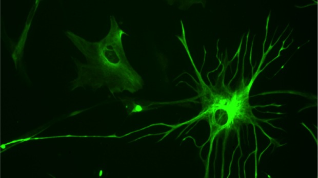
Other groups are working to convert reactive astrocytes—a form of glial ...

Interested in reading more?

Become a Member of

The Scientist Logo
Receive full access to more than 35 years of archives, as well as TS Digest, digital editions of The Scientist, feature stories, and much more!
Already a member? Login Here

Related Topics

Meet the Author

  • kerry grens

    Kerry served as The Scientist’s news director until 2021. Before joining The Scientist in 2013, she was a stringer for Reuters Health, the senior health and science reporter at WHYY in Philadelphia, and the health and science reporter at New Hampshire Public Radio. Kerry got her start in journalism as a AAAS Mass Media fellow at KUNC in Colorado. She has a master’s in biological sciences from Stanford University and a biology degree from Loyola University Chicago.

    View Full Profile
Share
Illustration of a developing fetus surrounded by a clear fluid with a subtle yellow tinge, representing amniotic fluid.
January 2026, Issue 1

What Is the Amniotic Fluid Composed of?

The liquid world of fetal development provides a rich source of nutrition and protection tailored to meet the needs of the growing fetus.

View this Issue
Skip the Wait for Protein Stability Data with Aunty

Skip the Wait for Protein Stability Data with Aunty

Unchained Labs
Graphic of three DNA helices in various colors

An Automated DNA-to-Data Framework for Production-Scale Sequencing

illumina
Exploring Cellular Organization with Spatial Proteomics

Exploring Cellular Organization with Spatial Proteomics

Abstract illustration of spheres with multiple layers, representing endoderm, ectoderm, and mesoderm derived organoids

Organoid Origins and How to Grow Them

Thermo Fisher Logo

Products

Brandtech Logo

BRANDTECH Scientific Introduces the Transferpette® pro Micropipette: A New Twist on Comfort and Control

Biotium Logo

Biotium Launches GlycoLiner™ Cell Surface Glycoprotein Labeling Kits for Rapid and Selective Cell Surface Imaging

Colorful abstract spiral dot pattern on a black background

Thermo Scientific X and S Series General Purpose Centrifuges

Thermo Fisher Logo
Abstract background with red and blue laser lights

VANTAstar Flexible microplate reader with simplified workflows

BMG LABTECH