Next Generation: Sneaking into a Cell

A nanoscale device measures electrical signals inside cells without causing damage.

Written byMegan Scudellari
| 3 min read

Register for free to listen to this article
Listen with Speechify
0:00
3:00
Share

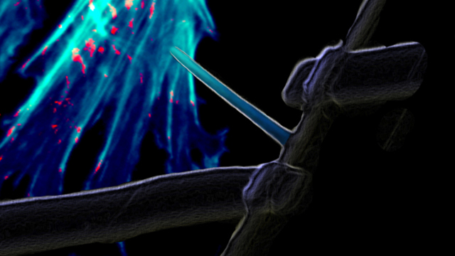
A false color SEM image of the BIT-FET overlaid with an image of a cluster of cardiomyocyte cells, illustrating how intracellular action potentials are recorded by the device L. ROBLES, OPUS DESIGN & X. DUAN, C.M LIEBER, HARVARD UNIVERSITY

THE DEVICE: It is not easy to record the electrical signals that pass fleetingly through neurons and cardiomyocytes. But with a novel nanoscale device developed by Charles Lieber and colleagues at Harvard University, scientists can record these action potentials without damaging cells and even probe sub-cellular structures like dendrites, according to a report published last month (December 18) in Nature Nanotechnology.

The branched intracellular nanotube field-effect transistor, or BIT-FET, joins a nanowire and a nanotube into a slim T-shaped structure that can be inserted into a cell up to five times in the same place without disrupting the action potential or damaging the cell. The tiny hollow nanotube, 50-100 nanometers wide, penetrates the cell, sucking up a bit of the cytosol as it enters. This ...

Interested in reading more?

Become a Member of

The Scientist Logo
Receive full access to more than 35 years of archives, as well as TS Digest, digital editions of The Scientist, feature stories, and much more!
Already a member? Login Here

Related Topics

Meet the Author

Share
Illustration of a developing fetus surrounded by a clear fluid with a subtle yellow tinge, representing amniotic fluid.
January 2026, Issue 1

What Is the Amniotic Fluid Composed of?

The liquid world of fetal development provides a rich source of nutrition and protection tailored to meet the needs of the growing fetus.

View this Issue
Skip the Wait for Protein Stability Data with Aunty

Skip the Wait for Protein Stability Data with Aunty

Unchained Labs
Graphic of three DNA helices in various colors

An Automated DNA-to-Data Framework for Production-Scale Sequencing

illumina
Exploring Cellular Organization with Spatial Proteomics

Exploring Cellular Organization with Spatial Proteomics

Abstract illustration of spheres with multiple layers, representing endoderm, ectoderm, and mesoderm derived organoids

Organoid Origins and How to Grow Them

Thermo Fisher Logo

Products

Brandtech Logo

BRANDTECH Scientific Introduces the Transferpette® pro Micropipette: A New Twist on Comfort and Control

Biotium Logo

Biotium Launches GlycoLiner™ Cell Surface Glycoprotein Labeling Kits for Rapid and Selective Cell Surface Imaging

Colorful abstract spiral dot pattern on a black background

Thermo Scientific X and S Series General Purpose Centrifuges

Thermo Fisher Logo
Abstract background with red and blue laser lights

VANTAstar Flexible microplate reader with simplified workflows

BMG LABTECH