Nipah Protein Structure Revealed

The structure of a key protein of the deadly virus could serve as a stepping stone to antiviral therapy.

Written byJef Akst
| 2 min read

Register for free to listen to this article
Listen with Speechify
0:00
2:00
Share

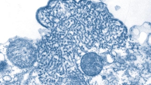
A transmission electron micrograph of Nipah virions isolated from a patient's cerebrospinal fluidWIKIMEDIA, CDCThe Nipah virus phosphoprotein, P, plays a key role in viral replication. Therefore, understanding its structure may shed light on ways to disrupt the viral life cycle. “If you can prevent the virus from making more RNA, then it can’t replicate, which is a good strategy for developing antiviral medications,” Jessica Bruhn, a graduate student in Erica Ollmann Saphire’s lab at Scripps Research Institute in La Jolla, California, said in a press release.

With this in mind, Bruhn, Saphire, and colleagues subjected the protein to X-ray crystallography, observing diffraction patterns that revealed its shape: “a long, parallel, tetrameric, coiled coil with a small, α-helical cap structure,” according the paper published in the December issue of the Journal of Virology. Interestingly, this shape is highly similar to the P protein structure of the sendai, measles, and mumps paramyxoviruses, all of which are long, tetrameric coils, despite the fact that these viruses share little sequence identity with one another or with Nipah.

“It was surprising to us that this structure is so similar to those from measles and mumps viruses, even though they are only 5 [percent] to 26 percent identical in sequence,” Bruhn said in the release. The results ...

Interested in reading more?

Become a Member of

The Scientist Logo
Receive full access to more than 35 years of archives, as well as TS Digest, digital editions of The Scientist, feature stories, and much more!
Already a member? Login Here

Related Topics

Meet the Author

  • Jef (an unusual nickname for Jennifer) got her master’s degree from Indiana University in April 2009 studying the mating behavior of seahorses. After four years of diving off the Gulf Coast of Tampa and performing behavioral experiments at the Tennessee Aquarium in Chattanooga, she left research to pursue a career in science writing. As The Scientist's managing editor, Jef edited features and oversaw the production of the TS Digest and quarterly print magazine. In 2022, her feature on uterus transplantation earned first place in the trade category of the Awards for Excellence in Health Care Journalism. She is a member of the National Association of Science Writers.

    View Full Profile
Share
Illustration of a developing fetus surrounded by a clear fluid with a subtle yellow tinge, representing amniotic fluid.
January 2026, Issue 1

What Is the Amniotic Fluid Composed of?

The liquid world of fetal development provides a rich source of nutrition and protection tailored to meet the needs of the growing fetus.

View this Issue
Skip the Wait for Protein Stability Data with Aunty

Skip the Wait for Protein Stability Data with Aunty

Unchained Labs
Graphic of three DNA helices in various colors

An Automated DNA-to-Data Framework for Production-Scale Sequencing

illumina
Exploring Cellular Organization with Spatial Proteomics

Exploring Cellular Organization with Spatial Proteomics

Abstract illustration of spheres with multiple layers, representing endoderm, ectoderm, and mesoderm derived organoids

Organoid Origins and How to Grow Them

Thermo Fisher Logo

Products

Brandtech Logo

BRANDTECH Scientific Introduces the Transferpette® pro Micropipette: A New Twist on Comfort and Control

Biotium Logo

Biotium Launches GlycoLiner™ Cell Surface Glycoprotein Labeling Kits for Rapid and Selective Cell Surface Imaging

Colorful abstract spiral dot pattern on a black background

Thermo Scientific X and S Series General Purpose Centrifuges

Thermo Fisher Logo
Abstract background with red and blue laser lights

VANTAstar Flexible microplate reader with simplified workflows

BMG LABTECH