Renowned Microscopist Dies

Michael Davidson, a microphotographer at Florida State University, has passed away at age 65.

Written byCatherine Offord
| 2 min read

Register for free to listen to this article
Listen with Speechify
0:00
2:00
Share

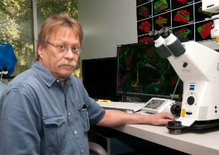
NATIONAL HIGH MAGNETIC FIELD LABORATORY/FLORIDA STATE UNIVERSITYMicroscopist Michael Davidson of Florida State University (FSU) in Tallahassee, whose images have appeared on everything from scientific papers to neckwear, died last month (December 24). He was 65.

During his time at the National High Magnetic Field Laboratory at FSU, Davidson and his team of more than 25 lab members (and 30 microscopes) developed several imaging techniques. In the mid-2000s, his collaboration with Eric Betzig and Harold Hess on photoactivated localization microscopy contributed to work that led to Betzig’s jointly receiving the Nobel Prize in Chemistry in 2014. “[Davidson] had a very big role in sparking the whole thing and enabling us to bring it to fruition,” Betzig told The New York Times this week (January 12).

Although never formally trained in art or photography, Davidson took stunning scientific images that landed him more than 1,500 magazine covers and—more unusually—decorated millions of neckties produced in partnership with Stonehenge Ltd. The ties, produced in ...

Interested in reading more?

Become a Member of

The Scientist Logo
Receive full access to more than 35 years of archives, as well as TS Digest, digital editions of The Scientist, feature stories, and much more!
Already a member? Login Here

Related Topics

Meet the Author

  • After undergraduate research with spiders at the University of Oxford and graduate research with ants at Princeton University, Catherine left arthropods and academia to become a science journalist. She has worked in various guises at The Scientist since 2016. As Senior Editor, she wrote articles for the online and print publications, and edited the magazine’s Notebook, Careers, and Bio Business sections. She reports on subjects ranging from cellular and molecular biology to research misconduct and science policy. Find more of her work at her website.

    View Full Profile
Share
Illustration of a developing fetus surrounded by a clear fluid with a subtle yellow tinge, representing amniotic fluid.
January 2026

What Is the Amniotic Fluid Composed of?

The liquid world of fetal development provides a rich source of nutrition and protection tailored to meet the needs of the growing fetus.

View this Issue
Redefining Immunology Through Advanced Technologies

Redefining Immunology Through Advanced Technologies

Ensuring Regulatory Compliance in AAV Manufacturing with Analytical Ultracentrifugation

Ensuring Regulatory Compliance in AAV Manufacturing with Analytical Ultracentrifugation

Beckman Coulter Logo
Skip the Wait for Protein Stability Data with Aunty

Skip the Wait for Protein Stability Data with Aunty

Unchained Labs
Graphic of three DNA helices in various colors

An Automated DNA-to-Data Framework for Production-Scale Sequencing

illumina

Products

nuclera logo

Nuclera eProtein Discovery System installed at leading Universities in Taiwan

Brandtech Logo

BRANDTECH Scientific Introduces the Transferpette® pro Micropipette: A New Twist on Comfort and Control

Biotium Logo

Biotium Launches GlycoLiner™ Cell Surface Glycoprotein Labeling Kits for Rapid and Selective Cell Surface Imaging

Colorful abstract spiral dot pattern on a black background

Thermo Scientific X and S Series General Purpose Centrifuges

Thermo Fisher Logo