

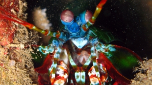
 A mantis shrimp in the waters surrounding IndonesiaWIKIMEDIA, SILKE BARONMantis shrimp have a unique view of the world. The crustaceans, along with other some insects and cephalopods, can sense difference in polarized light emissions, which arrive at an observer in one or more planes of direction. Mantis shrimp also use 16 different photoreceptor pigments, where mammals typically have two or three. Now, inspired by the marine organisms’ compound eyes and polarized vision, researchers have built a miniature sensor that can detect minute differences in early-stage cancerous cells that are invisible to the naked human eye. This enhanced contrast could be especially useful when paired with endoscopes to visually scan body regions, such as the colon, for developing cancers.
Reporting their work in this week’s Proceedings of the IEEE, an international team of researchers presented images of a cancerous mouse colon to demonstrate the device’s improvements over the bulkier, more complex, and expensive polarized imaging camera arrays already in existence. “We’ve moved from having multiple cameras to a single-chip solution,” Viktor Gruev, a computer scientist at Washington University in St. Louis and senior author on the paper, told Smithsonian. “It’s hard to put multiple cameras on an endoscope and take pictures. With our device, all the filters are on the camera and it goes from something that sits on your optical bench to one that goes on the end of an endoscope.”
While the technology will need to be refined and tested further before ...


