© SILVIA OTTE/GETTY IMAGES
James Black, a 62-year-old London taxi cab driver, went to his doctor complaining of memory difficulties and intermittent periods of confusion that he’d been experiencing for 2 years. A minor road accident caused by poor concentration and vision problems had forced him to retire. His wife reported that for more than a decade James had also experienced difficulty smelling—a condition, called hyposmia, that was confirmed by olfactory testing. His neurological examination revealed he was suffering from damage to the brain’s frontal lobe. Ultimately, James was diagnosed with Alzheimer’s disease (AD), the most common dementia-causing disorder.1
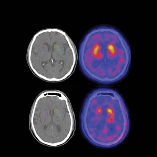
Importantly, some disorders commonly misdiagnosed as PD do not respond well to L-DOPA and other drugs that increase dopamine, a neurotransmitter involved in the control of motor function. Such agents are the most effective treatments available for PD patients. Thus, olfactory testing can aid physicians in predicting whether patients can derive meaningful benefit from such drugs. In patients with mild to moderate AD, olfactory testing indicates responsiveness to donepezil, a drug that improves cognitive function in some patients.2 In light of these and other findings, the Quality Standards Subcommittee of the American Academy of Neurology and other professional organizations have endorsed smell testing as an aid in the diagnosis of AD and PD. Nevertheless, the importance of olfaction in these diseases is largely overlooked, and such testing is not routinely performed in neurology clinics.
Predicting decline
Numerous studies have used quantitative smell tests in an attempt to identify asymptomatic older persons who are most likely to develop cognitive or motor symptoms indicative of neurodegenerative disease. In a pioneering study published in 1999, Amy Bornstein Graves and her associates at the University of South Florida administered a 12-item version of the University of Pennsylvania Smell Identification Test (UPSIT), termed the B-SIT, to 1,604 community-dwelling senior citizens who showed no signs of dementia.3 Over the course of the two-year study, the olfactory test scores proved to be a better predictor of cognitive decline than scores on a global cognitive test. Overall, individuals who had no sense of smell and who possessed at least one APOE-4 allele—a genetic risk factor for AD—were nearly five times more likely to develop cognitive decline than those of the same age who had no smell dysfunction and who carried no such allele. This risk was increased nearly tenfold in women, whereas in men it went up approximately threefold. Possessing at least one APOE-4 allele in the absence of smell loss did not significantly increase a person’s risk for future cognitive decline.
Evaluation of olfactory ability can help ensure the correct diagnosis and treatment strategy for neurodegenerative disease. Nevertheless, the importance of olfaction is largely overlooked, and such testing is not routinely performed
in neurology clinics.
A more recent study of 1,092 older persons with no signs of dementia (average age 80 years) from a multiethnic community in New York City also observed an association between smell loss and cognitive function. Those individuals with both mild cognitive impairment (MCI) and memory loss had lower scores on the 40-odor UPSIT than those with MCI but no memory loss. The UPSIT scores were also correlated with age, several cognitive measures, and the volume of the hippocampus, a brain structure associated with memory.?4
Research has also elucidated a link between smell dysfunction and PD. In the 1990s, G. Webster Ross and his colleagues at the University of Hawaii administered the B-SIT to 2,276 nonsymptomatic elderly men of Japanese ancestry (average age at the beginning of the study was 80 years). After adjusting for age, smoking behavior, and other confounders, those subjects whose initial olfactory test scores fell within the bottom 25 percent of the group were five times more likely to develop PD than those whose test scores fell within the top 25 percent. Over a four-year period, 35 were clinically diagnosed with PD.5
Further support for olfactory involvement in PD came in 2004, when Mirthe Ponsen and her associates at Vrije Universiteit in Amsterdam published a study of 361 asymptomatic first-degree relatives of PD patients, finding that those whose olfactory test scores were significantly below normal were more likely to develop PD over a two-year period than those with no smell impairment.6
Additionally, Ponsen measured the extent of degeneration of brain regions associated with PD-related motor dysfunction. The team injected patients with a radioactively labelled agent that binds to the dopamine transporter responsible for moving dopamine back into neurons in the brain following its release into the synaptic cleft, then measured the amount of such binding using gamma-ray cameras—a technique known as single-photon-emission computerized tomography (SPECT). The less binding detected, the greater the damage in the cells of interest.
At the two-year assessment, four of the 40 relatives with the lowest olfactory test scores—all of whom exhibited substantial reduction in the amount of dopamine transporter binding at the start of the study—were diagnosed with clinical PD, while none of the 38 relatives with the highest olfactory test scores developed the disease. After five years, those relatives with initial smell loss had at least a 10 percent risk of developing clinically defined PD.7 When the degeneration in the brain regions producing dopamine as measured by SPECT were taken into account, the investigators suggested that the risk of developing PD in the presence of hyposmia may be as high as 22 percent.
Such studies suggest that olfactory testing can sometimes predict future development of cognitive and, in the case of PD, motor dysfunction in those at risk for degenerative disease. Although the predictive power of olfactory testing alone is not high, it rivals and even exceeds that of a number of other biomarkers of PD, including disease-related metabolites found in the cerebral spinal fluid and SPECT imaging of the dopamine transporter. Importantly, novel methods are being developed that enhance smell testing’s predictive power. For example, intranasal application of atropine, a drug that accentuates cognitive dysfunction in patients with AD, may induce a greater degree of smell loss in symptomless individuals who are at risk for future dementia—in effect “unmasking” the incipient disease.8
The root of the problem
© TAMI TOLPAWhile smell dysfunction can be useful in differential diagnosis, the fact remains that many neurological diseases exhibit essentially equivalent olfactory loss, including disorders with neuropathologies completely distinct from those of AD and PD, such as myasthenia gravis, an autoimmune disorder characterized by muscle weakness.9 Hence, while olfactory dysfunction is a sensitive indicator of some neurological diseases, it is not specific to any single disease. Is it possible that the same pathological process is involved in the olfactory loss associated with most or all of these disorders, or is the olfactory system simply sensitive to damage from a range of disease-specific factors?
One hypothesis is that deficits in certain neurotransmitter systems are largely responsible for smell dysfunction that occurs in conjunction with neurodegenerative disease. A major player in this regard is the basal forebrain cholinergic system, which is involved in the secretion of the neurotransmitter acetylcholine (ACh) in many areas of the brain. ACh plays a significant role in attention, memory, and the facilitation of cortical plasticity, including functional recovery following brain injury.
Cholinergic neurons that project from the basal forebrain to the olfactory bulb also directly modulate neural activity and inhibit the activity of cells critical for immune responses to brain damage and foreign agents, including microglia, astrocytes, and oligodendrocytes.10 When cholinergic and other neural cells that project to the olfactory bulb are damaged, inhibition of microglia can be suppressed, resulting in immune activation and the secretion of inflammatory mediators such as the cytokine tumor necrosis factor–alpha (TNF–α). (See illustration here.) Although low levels of TNF–α are neuroprotective, high levels induce damaging inflammation and even cell death. Activated glial cells and local inflammatory processes are believed to contribute to the development of a number of neurodegenerative diseases, and may also contribute to the degeneration of the basal forebrain cholinergic system, resulting in olfactory impairment.
Several lines of evidence support the concept that cholinergic dysfunction plays a significant role in the olfactory loss seen in a number of neurological diseases.
First, the ability of patients with PD to identify odors has recently been shown to be correlated with the degree of cholinergic denervation of the forebrain, as measured by functional imaging of radioactively labeled agents that bind to acetylcholine receptors.11
COURTESY OF SENSONICSSecond, autopsy studies show that disorders with an olfactory dysfunction element are typically accompanied by significant damage to the forebrain cholinergic structures. Such damage, which in most cases is reflected by gliosis (the glial response to brain damage) and cell loss, is less severe or absent in diseases whose olfactory function is spared or less compromised, including depression and essential tremor. Although cell loss within basal forebrain cholinergic structures is minimal in patients with Huntington’s disease, another disorder with marked olfactory loss, their cholinergic system is nonetheless dysfunctional. Specifically, patients exhibit changes in the expression of choline acetyltransferase (ChAT), which is involved in ACh synthesis, and of vesicular acetylcholine transporter, a protein critical for transporting ACh from the cytoplasm into the synaptic vesicles.12
Third, another measure of cholinergic circuit health, called short-latency afferent inhibition (SAI), also correlates with relative olfactory function differences in patients with neurodegenerative disease.13 SAI is measured by electrically stimulating a sensory nerve in the hand immediately before activating the motor cortex by transcranial magnetic stimulation (TMS), a noninvasive procedure in which magnetic coils on the surface of the scalp are used to stimulate cortical neurons. When electrical stimulation is applied to the sensory nerve just before the onset of TMS, the subsequent activation of motor neurons in the muscles of the arm is delayed or inhibited. In AD and PD, particularly PD with dementia, such inhibition is less marked.
Finally, a large literature based on animal studies clearly links olfactory function to cholinergic processes. For example, mouse strains that express fewer α7-nicotinic cholinergic receptors perform poorly on odor detection/discrimination tasks relative to strains that have more of these receptors. Other studies have shown that physostigmine, a drug that inhibits an enzyme that decreases the amount of acetylcholine at synapses, increases the ability of rats to discriminate between low concentrations of odors.
Much remains to be learned about the factors that initiate cholinergic dysfunction and the degree to which dysregulation of noncholinergic neuromodulators, such as serotonin and norepinephrine, contributes to olfactory loss. For example, in diseases associated with abnormal aggregates of α-synuclein, tau, and β-amyloid (Aβ), it is unclear whether the olfactory deficits precede or follow the development of such neuropathology. Relatively strong correlations have been found between olfactory test scores very late in life and the number of such pathological structures in the postmortem brains of both healthy older persons and in older persons with AD, PD, and Lewy body disease.
Additionally, various steps in acetylcholine synthesis and release can be altered by Aβ-related peptides associated with AD. For example, exposure of brain slices from the hippocampus and cortex of rats to very low concentrations of Aβ and Aβ fragments can inhibit potassium-evoked release of acetylcholine. This does not occur in brain slices from the rat striatum, suggesting regional selectivity of such effects. Researchers have also shown that Lewy bodies, the defining α-synuclein-comprised pathological features of PD and Lewy body dementia, sequester precursor enzymes that are critical for the expression of acetylcholine and some other neurotransmitters, presumably disrupting neurotransmitter production.14 Whether these aggregates are contributing to olfactory dysfunction remains to be determined.
Environment and behavior
Many environmental and behavioral risk factors for a number of neurodegenerative diseases are also risk factors for smell dysfunction. For example, age is a risk factor for AD and PD, and smell loss is common in healthy older persons. Furthermore, viral and bacterial infections, notably those of the upper respiratory tract, are the most frequent cause of chronic, often permanent, smell loss in the general population, and a number of viruses and bacteria have been indirectly implicated in the etiology of neurodegenerative diseases.15 Decreased smell function can also result from head trauma and chronic exposure to ionized metals and air pollution, known risk factors for AD and PD.
It is well established that airborne toxins, viruses, nanoparticles, and other foreign substances—collectively called xenobiotics—can enter the brain through the nose via the olfactory epithelium, either damaging receptor cells directly or initiating harmful inflammatory responses, ultimately altering olfactory function.16,17 The olfactory epithelium is protected to a large degree by detoxification enzymes, including some encoded by members of the P450 gene superfamily. However, protection provided by such detoxifying enzymes—which in some instances are found at higher levels in the nose than in the liver—can be compromised. Postmortem studies have identified nanoparticles and inflammatory mediators in the olfactory epithelia and bulbs of children and young adults living in highly polluted areas of Mexico City.17 In some cases, AD- and PD-like pathology has been observed in their brains. Many young people living in these highly polluted areas also exhibit subtle olfactory dysfunction. Furthermore, iron deficiency enhances the uptake of manganese through the olfactory epithelium in rats, and in humans, anemia has been found to be a risk factor for both AD and PD.
Is it possible that the same pathological process is involved in the olfactory loss associated with most or all neurodegenerative disorders, or is the olfactory system simply sensitive to damage from a range of disease-specific factors?
It is not beyond the realm of possibility that damage to neurotransmitter systems from pathogens or other xenobiotic agents could be instrumental in altering basic metabolic and immunological activity in numerous brain regions. Such damage might then cause, catalyze, or hasten the formation of Lewy bodies, neuritic plaques, neurofibrillary tangles, and other pathologic entities that, in turn, alter the functioning of cells within the olfactory pathways. It is also possible that such damage, which might alter smell function, exacerbates nascent or latent disease-related brain pathology that otherwise would not be expressed.16
Ultimately, the expression of most neurodegenerative disease neuropathology and symptoms, including olfactory dysfunction, depends on myriad factors involving health, genetic predispositions, sex, age, and environmental exposure to disease-related risk factors. The complexity of neurodegenerative pathologies—which often span a number of diseases—remains a challenge not only for the diagnostician, but also for those seeking to develop treatments that target elements of such pathologies.
Clearly, future research is needed to better define the connection between olfaction and the pathologic processes associated with neurodegenerative disease. Is olfactory dysfunction a result of damage associated with classic markers of neurodegenerative disease, such as abnormal aggregates of α-synuclein, tau, and Aβ? Or does olfactory loss precede such damage? Can damage to the olfactory system, per se, induce neurodegenerative disease in those genetically or otherwise predisposed to such disease? These and a host of other questions await clarification.
Richard L. Doty is the director of the Smell and Taste Center and a professor in the Department of Otorhinolaryngology at the University of Pennsylvania’s Perelman School of Medicine. He is also president of Sensonics International, a corporation that manufactures smell and taste tests.
References
- C.H. Hawkes, R.L. Doty, The Neurology of Olfaction (Cambridge, UK: Cambridge University Press, 2009), 159.
- L. Velayudhan, S. Lovestone, “Smell identification test as a treatment response marker in patients with Alzheimer disease receiving donepezil,” J Clin Psychopharmacol, 29:387-90, 2009.
- A.B. Graves et al., “Impaired olfaction as a marker for cognitive decline: interaction with apolipoprotein E epsilon4 status,” Neurology, 53:1480-87, 1999.
- D.P. Devanand et al., “Olfactory identification deficits and MCI in a multi-ethnic elderly community sample,” Neurobiol Aging, 31:1593-600, 2010.
- G.W. Ross et al., “Association of olfactory dysfunction with risk for future Parkinson’s disease,” Ann Neurol, 63:167-73, 2008.
- M.M. Ponsen et al., “Idiopathic hyposmia as a preclinical sign of Parkinson’s disease,” Ann Neurol, 56:173-81, 2004.
- M.M. Ponsen et al., “Olfactory testing combined with dopamine transporter imaging as a method to detect prodromal Parkinson’s disease,” J Neurol Neurosurg Psychiatry, 81:396-99, 2010.
- P.W. Schofield et al., “An olfactory ‘stress test’ may detect preclinical Alzheimer’s disease,” BMC Neurol, 12:24, 2012.
- F.E. Leon-Sarmiento et al., “Profound olfactory dysfunction in myasthenia gravis,” PLOS One, 7:e45544, 2012.
- R.L. Doty, “Olfaction in Parkinson’s disease and related disorders,” Neurobiol Dis, 46:527-52, 2012.
- N.I. Bohnen et al., “Olfactory dysfunction, central cholinergic integrity and cognitive impairment in Parkinson’s disease,” Brain, 133:1747-54, 2010.
- R. Smith et al., “Cholinergic neuronal defect without cell loss in Huntington’s disease,” Hum Mol Genet, 15:3119-31, 2006.
- R. Nardone et al., “Cholinergic cortical circuits in Parkinson’s disease and in progressive supranuclear palsy: a transcranial magnetic stimulation study,” Exp Brain Res, 163:128-31, 2005.
- B.N. Dugger, D.W. Dickson, “Cell type specific sequestration of choline acetyltransferase and tyrosine hydroxylase within Lewy bodies,” Acta Neuropathol, 120:633-39, 2010.
- F. Mawanda, R. Wallace, “Can infections cause Alzheimer’s disease?” Epidemiol Rev, 35:161-80, 2013.
- R.L. Doty, “The olfactory vector hypothesis of neurodegenerative disease: is it viable?” Ann Neurol, 63:7-15, 2008.
- L. Calderón-Garcidueñas et al., “Urban air pollution: Influences on olfactory function and pathology in exposed children and young adults,” Exp Toxicol Pathol, 62:91-102, 2010.