
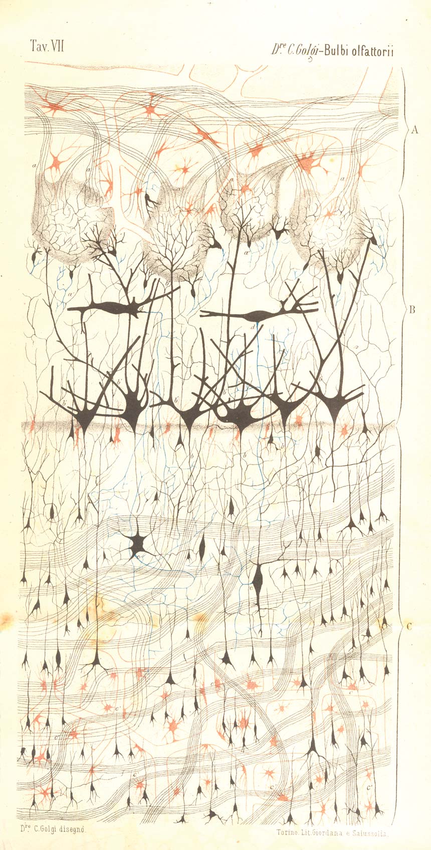
 A FIRST LOOK: Camillo Golgi’s original black-and-white drawing of a dog’s olfactory bulb appeared in a paper in the Rivista Sperimentale di Freniatria e Medicina Legale in 1875. Colored plates appeared in reprinted versions distributed by Golgi and in the German translation of his paper. Golgi indicated three layers in his drawing. The superficial layer (A) is composed of nerve fiber bundles made of myelinated axons (black lines), a branching blood vessel (outlined in red), and glial cells (red). The nerve fiber bundles enter spheres of cell bodies called glomeruli in the middle layer (B), which consists of gray matter (nerve cell bodies and dendrites). Large mitral cells lining the middle layer (black) are arranged such that their dendrites reach toward the glomeruli, where they form synapses with the axons extending from the superficial layer. The mitral cell axons (some of which are shown in blue) aim for the inner layer (C) of the olfactory bulb. The inner layer also contains granule cells, blood vessels, glial cells, and nerve fibers.
See larger image: JPGCOURTESY OF PAOLO MAZZARELLO, SISTEMA MUSEALE DI ATENEO, PAVIA, ITALYIt’s 1873 at a hospice hospital near Milan, and a young Italian physician named Camillo Golgi is surrounded by brains and nervous tissue from animals including cows and dogs, as well as from recently departed patients. Unaware that he will one day become famous for, among many things, discovering the “Golgi body,” the 30-year-old scientist currently has a single goal: to capture the mysterious details of neurons by staining them with a concoction of his own design.
He impregnates formalin-fixed nervous tissue sections with potassium dichromate and silver nitrate, causing microcrystals of silver chromate to fill in the delicate dendrites and axons of neurons. The technique paints the structures black and makes them visible in situ for the very first time. Golgi would spend several years perfecting the reaction, which likely started out as an accidental discovery. According to Paolo Mazzarello, director of the Golgi Museum in Pavia, Italy, Golgi may have first noticed the reaction’s potential to mark neurons while staining connective tissues surrounding the brain’s blood vessels with silver nitrate, a commonly used compound at the time. “He probably saw some partial staining of the [nerve] cells,” and from there, a combination of chance and intuition led Golgi to develop ...


