
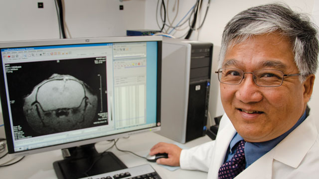
 Philip Liu with a high-resolution MRI image demonstrating gene transcription in a mouse brain using RNA labeled with superparamagnetic iron oxide nanoparticles (dark areas indicate the presence of the RNA of interest). LAURIE LIZOTTE/REMS MEDIA SERVICES
Neuroimaging isn’t just about pretty pictures anymore. Microscopists and other imaging scientists are now looking beyond mere anatomy, gleaning vital information about in vivo brain activity from their digital films. They can see where blood flow speeds up, when nerves fire, and what genes are switched on in a cell—all, crucially, without resorting to slicing up the brain.
In the 21st-century neuroimaging lab, mice are running around with lightweight microscopes mounted to their craniums. Scientists are using advanced techniques that rely on natural contrast instead of fluorescent markers. They are revamping an old method, ultrasound, to obtain detailed images of brain blood flow. Furthermore, researchers are combining molecular tools with standard imaging modes in new ways to track cells’ behavior, scalpel-free.
Driving the development of ...


