

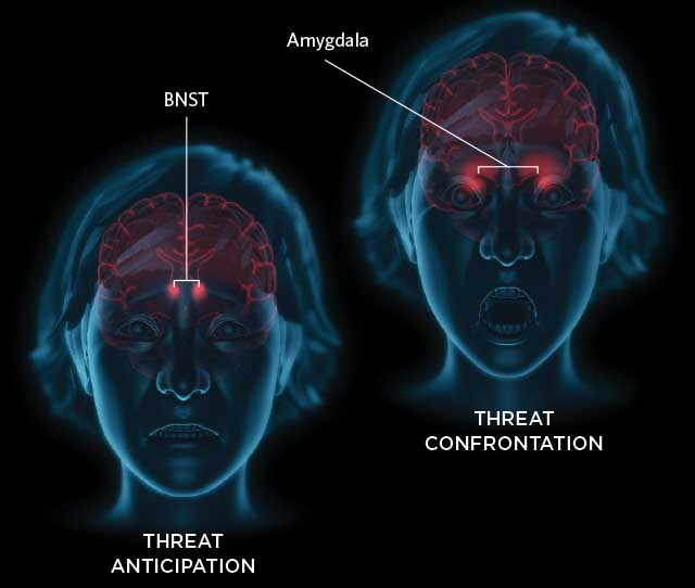
 ON EDGE: Researchers trained participants in two studies to associate visual cues with a mild electric shock to the finger. Following a visual cue suggesting a shock might be imminent—i.e., during threat anticipation—the volunteers’ brains showed higher activity in the bed nucleus of the stria terminalis (BNST), a region of the brain associated with defensive responses in uncertain situations. When participants were shocked—i.e., during threat confrontation—they showed higher activity in their amygdalae, two almond-shaped clusters of nuclei associated with fear and emotional stimulation.© EVAN OTO/SCIENCE SOURCE
The paper
F. Klumpers et al., “How human amygdala and bed nucleus of the stria terminalis may drive distinct defensive responses,” J Neurosci, 37:9645-56, 2017.
When Floris Klumpers zapped people with electricity while working toward his PhD in the late 2000s, he expected his volunteers’ amygdalae—key emotion centers in the brain—to light up in anticipation of a shock. “There was this idea that the amygdala is the most important structure in emotion processing—especially in fear processing,” says Klumpers, then at Utrecht University in the Netherlands. “We were quite surprised, using fMRI studies, to not find amygdala activity when people were anticipating an adverse event.”
Klumpers assumed he’d made a mistake, but after replicating the finding in further experimental work, he began thinking about the different ...


