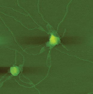
 NEW ANATOMY: Long nanofilaments extend from two glioblastoma-derived exosomes.COURTESY SHIVANI SHARMA
The paper
S. Sharma et al., “Nanofilaments on glioblastoma exosomes revealed by peak force microscopy,” J Royal Soc Interface, doi:10.1098/rsif.2013.1150, 2014.
The approach
Exosomes are ball-shaped, secreted vesicles involved in intercellular chatter, including the delivery of metastatic messages. (See “Exosome Explosion,” The Scientist, July 2011.) Given exosomes’ tiny size—roughly 100 nm in diameter—electron microscopy has limited how much scientists can resolve of their structure, says Shivani Sharma of the University of California, Los Angeles. So she and her colleagues turned to peak force microscopy, a variant of atomic force microscopy, which can “feel” the shape of objects at the nanoscale.
The structure The team observed filaments, about 10–20 nm wide and up to several microns in length, protruding from glioblastoma-derived exosomes, but not from exosomes ...


