Intracellular Spirals

Membrane twists connect stacked endoplasmic reticulum sheets.

Written byKate Yandell
| 2 min read

Register for free to listen to this article
Listen with Speechify
0:00
2:00
Share

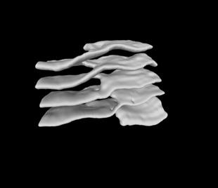
RAMP IT UP: Sheets of endoplasmic reticulum are connected by a twist of membrane such that the entire structure resembles the spiral of a parking garage.CELL, TERASAKI ET AL.

The paper M. Terasaki et al., “Stacked endoplasmic reticulum sheets are connected by helicoidal membrane motifs,” Cell, 154:285-96, 2013. The finding The endoplasmic reticulum (ER), an organelle composed of interconnected sheets and tubules, is well known as a site of protein synthesis. But the fine structure of its characteristic folds, and in particular how they connect, is poorly understood. Researchers now show that ER sheets in mouse salivary gland and neuronal cells are connected to their neighbors by twists of membrane, and the stack of sheets forms a spirally connected structure resembling a parking garage. The method In the past, researchers have tended to look at isolated cross sections of stacked ER. Instead, Mark Terasaki, a cell biologist at the University of Connecticut Health Center, ...

Interested in reading more?

Become a Member of

The Scientist Logo
Receive full access to digital editions of The Scientist, as well as TS Digest, feature stories, more than 35 years of archives, and much more!
Already a member? Login Here
Illustration of a developing fetus surrounded by a clear fluid with a subtle yellow tinge, representing amniotic fluid.
January 2026, Issue 1

What Is the Amniotic Fluid Composed of?

The liquid world of fetal development provides a rich source of nutrition and protection tailored to meet the needs of the growing fetus.

View this Issue
Skip the Wait for Protein Stability Data with Aunty

Skip the Wait for Protein Stability Data with Aunty

Unchained Labs
Graphic of three DNA helices in various colors

An Automated DNA-to-Data Framework for Production-Scale Sequencing

illumina
Exploring Cellular Organization with Spatial Proteomics

Exploring Cellular Organization with Spatial Proteomics

Abstract illustration of spheres with multiple layers, representing endoderm, ectoderm, and mesoderm derived organoids

Organoid Origins and How to Grow Them

Thermo Fisher Logo

Products

Brandtech Logo

BRANDTECH Scientific Introduces the Transferpette® pro Micropipette: A New Twist on Comfort and Control

Biotium Logo

Biotium Launches GlycoLiner™ Cell Surface Glycoprotein Labeling Kits for Rapid and Selective Cell Surface Imaging

Colorful abstract spiral dot pattern on a black background

Thermo Scientific X and S Series General Purpose Centrifuges

Thermo Fisher Logo
Abstract background with red and blue laser lights

VANTAstar Flexible microplate reader with simplified workflows

BMG LABTECH