Neural Connectome Method Uses mRNA Barcodes

Researchers swap microscopy for RNA sequencing to track neural paths in the mouse brain.

Written byRuth Williams
| 3 min read

Register for free to listen to this article
Listen with Speechify
0:00
3:00
Share

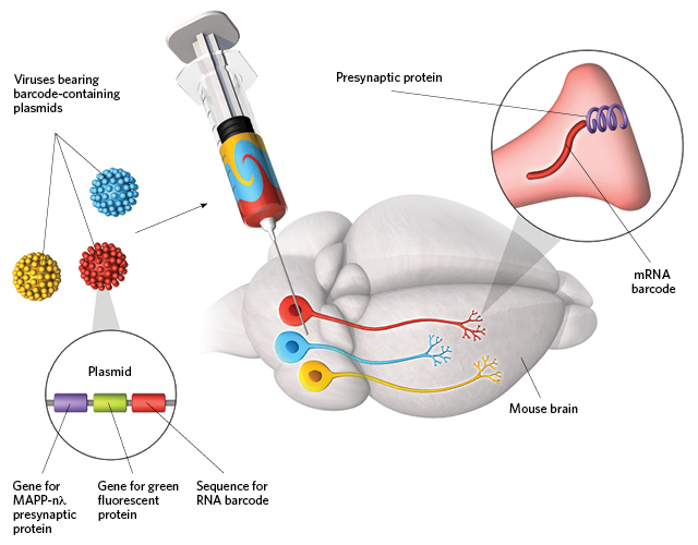
FOLLOW THE mRNA: To determine where in the mouse brain individual neurons have axon terminals, researchers inject a library of viral vectors in the vicinity of cell bodies. The viruses contain plasmids each encoding a unique RNA barcode and a presynaptic protein called MAPP-nλ that will shuttle the barcoded mRNA to the ends of the axon. Typically, each cell takes up only one virus, giving that cell an RNA identifier. Dissecting brain regions and sequencing the RNAs reveals which cells project where.© GEORGE RETSECK

Several ambitious brain connectome projects are underway to diagram the wiring of animals’ most complicated organ. For the most part, neural cartographers rely on microscopy, but tracing numerous projections across the large volumes of the brain is “extremely painstaking and slow,” says Thomas Mrsic-Flogel of the University of Basel.

Inspired by recent advances in sequencing technology, Tony Zador of Cold Spring Harbor Laboratory had an idea: “If there was a way to convert the mapping problem into one of sequencing, it would be a [big] win in terms of throughput and cost.” Now, he and his colleagues have done just that.

To map neurons by sequencing, Zador’s team injects a mixture of approximately 106 viruses—each bearing a genetic sequence encoding a unique mRNA barcode—into mouse ...

Interested in reading more?

Become a Member of

The Scientist Logo
Receive full access to digital editions of The Scientist, as well as TS Digest, feature stories, more than 35 years of archives, and much more!
Already a member? Login Here

Related Topics

Meet the Author

  • ruth williams

    Ruth is a freelance journalist. Before freelancing, Ruth was a news editor for the Journal of Cell Biology in New York and an assistant editor for Nature Reviews Neuroscience in London. Prior to that, she was a bona fide pipette-wielding, test tube–shaking, lab coat–shirking research scientist. She has a PhD in genetics from King’s College London, and was a postdoc in stem cell biology at Imperial College London. Today she lives and writes in Connecticut.

    View Full Profile

Published In

November 2016

Nimble Neurons

The remarkable adaptability of the nervous system

Share
December digest cover image of a wooden sculpture comprised of multiple wooden neurons that form a seahorse.
December 2025, Issue 1

Wooden Neurons: An Artistic Vision of the Brain

A neurobiologist, who loves the morphology of cells, turns these shapes into works of art made from wood.

View this Issue
Stacks of cell culture dishes, plates, and flasks with pink cell culture medium on a white background.

Driving Innovation with Cell Culture Essentials

Merck
Stacks of cell culture dishes, plates, and flasks with pink cell culture medium on a white background.

Driving Innovation with Cell Culture Essentials

MilliporeSigma purple logo
Abstract wireframe sphere with colorful dots and connecting lines representing the complex cellular and molecular interactions within the tumor microenvironment.

Exploring the Inflammatory Tumor Microenvironment 

Cellecta logo
An image of a DNA sequencing spectrum with a radial blur filter applied.

A Comprehensive Guide to Next-Generation Sequencing

Integra Logo

Products

brandtech logo

BRANDTECH® Scientific Announces Strategic Partnership with Copia Scientific to Strengthen Sales and Service of the BRAND® Liquid Handling Station (LHS) 

Top Innovations 2026 Contest Image

Enter Our 2026 Top Innovations Contest

Biotium Logo

Biotium Expands Tyramide Signal Amplification Portfolio with Brighter and More Stable Dyes for Enhanced Spatial Imaging

Labvantage Logo

LabVantage Solutions Awarded $22.3 Million U.S Customs and Border Protection Contract to Deliver Next-Generation Forensic LIMS