New Tests for Zika in the Works

To answer questions about the risks of Zika virus infection, researchers need better diagnostics.

Written byKerry Grens
| 3 min read

Register for free to listen to this article
Listen with Speechify
0:00
3:00
Share

WIKIMEDIA, JEFFREY M. VINOCURAs reports of Zika virus infections continue to spread through the Americas, countless questions loom. Chief among them is about the relationship between infection during pregnancy and microcephaly in babies, which has been difficult to pin down given the limitations of current diagnostics. A number of researchers are working at breakneck speed to develop immunological reagents and assays that could confirm whether a person has had a Zika infection.

“We’re trying to do the best we can to give some answers to the clinicians relatively soon,” said Nikos Vasilakis, who is developing Zika tests at the University of Texas Medical Branch in Galveston.

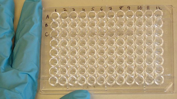
Currently, the standard assay for Zika viral infection is a PCR test that probes for the presence of viral RNA in a sample. While it works well to detect the virus, the pathogen’s RNA is only around for a short period of time. “By the time [patients] make it into the clinic, the virus is likely gone or it’s at the tail end, beyond the limit of detection,” said Vasilakis.

What clinicians and epidemiologists would really like is to be able to determine whether a baby was exposed to Zika in ...

Interested in reading more?

Become a Member of

The Scientist Logo
Receive full access to more than 35 years of archives, as well as TS Digest, digital editions of The Scientist, feature stories, and much more!
Already a member? Login Here

Related Topics

Meet the Author

  • kerry grens

    Kerry served as The Scientist’s news director until 2021. Before joining The Scientist in 2013, she was a stringer for Reuters Health, the senior health and science reporter at WHYY in Philadelphia, and the health and science reporter at New Hampshire Public Radio. Kerry got her start in journalism as a AAAS Mass Media fellow at KUNC in Colorado. She has a master’s in biological sciences from Stanford University and a biology degree from Loyola University Chicago.

    View Full Profile
Share
Illustration of a developing fetus surrounded by a clear fluid with a subtle yellow tinge, representing amniotic fluid.
January 2026, Issue 1

What Is the Amniotic Fluid Composed of?

The liquid world of fetal development provides a rich source of nutrition and protection tailored to meet the needs of the growing fetus.

View this Issue
Skip the Wait for Protein Stability Data with Aunty

Skip the Wait for Protein Stability Data with Aunty

Unchained Labs
Graphic of three DNA helices in various colors

An Automated DNA-to-Data Framework for Production-Scale Sequencing

illumina
Exploring Cellular Organization with Spatial Proteomics

Exploring Cellular Organization with Spatial Proteomics

Abstract illustration of spheres with multiple layers, representing endoderm, ectoderm, and mesoderm derived organoids

Organoid Origins and How to Grow Them

Thermo Fisher Logo

Products

Brandtech Logo

BRANDTECH Scientific Introduces the Transferpette® pro Micropipette: A New Twist on Comfort and Control

Biotium Logo

Biotium Launches GlycoLiner™ Cell Surface Glycoprotein Labeling Kits for Rapid and Selective Cell Surface Imaging

Colorful abstract spiral dot pattern on a black background

Thermo Scientific X and S Series General Purpose Centrifuges

Thermo Fisher Logo
Abstract background with red and blue laser lights

VANTAstar Flexible microplate reader with simplified workflows

BMG LABTECH