Microplate readers are widely used in research and industrial settings to examine biological, physical, and chemical reactions. These instruments work by detecting light signals from fluorescent and bioluminescent emitters or measuring how much light is absorbed or scattered as it passes through a sample. Microplate reader output is affected by several settings and parameters, and operators should understand how these features impact data consistency and accuracy.
Understanding Gain
The gain setting defines how a microplate reader amplifies incoming light signals. High gain enhances the visibility of dim signals against background noise, while low gain prevents oversaturation from bright signals. Modern plate readers can identify the optimal gain value based on standard curve readings. Users can then adjust this based on their sample well results. In general, gain adjustment is performed with a target value of 90 percent of the sample well expected to generate the strongest light signal. For kinetic measurements where light signal is expected to change over time, a target value of 10 percent of a blank sample well is recommended instead. Finally, the Enhanced Dynamic Range (EDR) function—found on VANTAstar®, PHERAstar® FSX, and CLARIOstar® Plus readers from BMG LABTECH—can continuously and automatically adjust gain settings throughout a measurement.

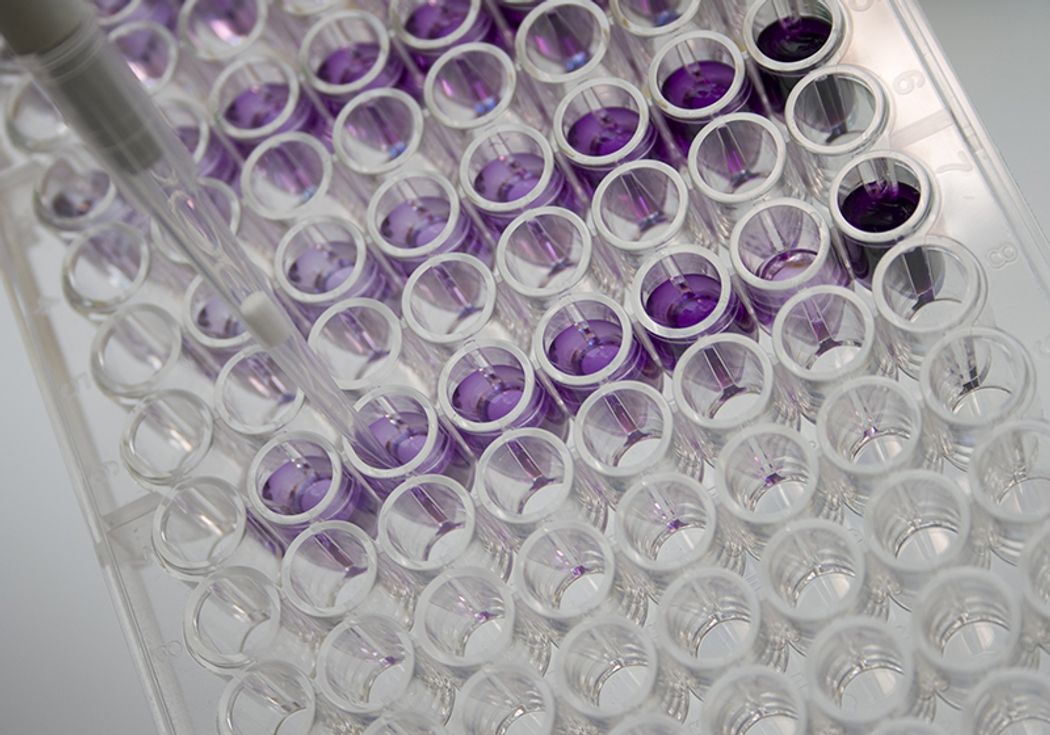
Microplate readers are used for numerous assays in biological research and industry.
iStock, scottiefone
Avoiding Autofluorescence
Cell-based assays preserve physiological context, giving researchers data with more biological relevance compared to assays using liquid solution-based samples. However, they also require additional considerations to mitigate increased signal noise and variability. Autofluorescence can be a significant problem when working with live cells, as both cellular elements and culture media components can produce nonspecific signals. Specifically, aromatic side chains, commonly found on carbohydrates, amino acids, proteins, and other elements essential to cell function and survival, emit signals in the blue and green portions of the spectrum. Researchers may circumvent or mitigate this to some degree by using red-shifted fluorophores, optically transparent media, and by avoiding the blue-green region for detection.
Finding Focus
Signal intensity is generally not uniform throughout a sample, especially when working with heterogeneous cell-based samples rather than homogenous liquid solutions. In liquid samples, the highest signal intensity is usually at a point slightly underneath the liquid surface. Conversely, the highest intensity will be at the bottom of the well when working with adherent cells. Here, reading from the bottom rather than the top can improve signal acquisition. In either situation, accurate and consistent fill volume can help reduce variability between samples, as a discrepancy of as little as a few microliters can lead to significant differences in measured signal intensity.
Microplate readers can have fixed, manually adjustable, or automatically adjustable focal heights. Instruments with fixed focal heights offer limited sensitivity, while devices with manual settings require some trial and error on the part of the user to find the proper setting. In contrast, readers capable of automated focus adjustments can determine the optimal settings prior to each experimental run based on a user-defined sample or group of samples.
The Meniscus Effect
A liquid stored in a container will typically form a meniscus—a concave or convex curvature in the upper surface caused by capillary action. In absorbance measurements, this changes the path length—the distance that light must travel through the sample from the emitting device to the sensor—creating discrepancies depending on where reads are taken within the well. Microplate samples usually form concave menisci, leading to shorter path lengths in the center of the well and longer ones at the edges.
Limiting the amount of buffers or detergents in the sample can mitigate the increased meniscus effect caused by these agents. Similarly, using untreated polystyrene microplates can limit meniscus formation because they are hydrophobic while microplates treated for tissue culture applications tend to become hydrophilic. Microplate geometry also affects meniscus formation. Square wells create less uniform meniscus formation, while U- or V-shaped wells create different path lengths even without factoring in any meniscus. Finally, filling wells to the brim eliminates the meniscus altogether. However, this can only be applied in certain situations where sample scarcity is not a factor.
Microplate reader instruments may offer path length correction, which uses water’s natural absorbance peak at ~970 nm to determine sample path length. This technique compensates for meniscus formation and can normalize for fill volume, but it can only be applied to aqueous solutions because it is established using water.
Optimizing Microplate Reader Data
Microplate readers are versatile instruments, but different assays will require different settings. Recognizing how instrument features can affect reader output can help scientists obtain more robust, accurate, and consistent data from their experiments.


