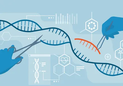
Molecular biologist and Nobel Prize winner Sydney Brenner notably said, “Progress in science depends on new technologies, new ideas, and new discoveries—in that order.”1 The field of gene therapy is a testament to Brenner’s point. Some of the most prevalent genetic diseases on Earth, such as sickle cell disease (SCD) and thalassemia, were discovered to have a molecular cause in the 1950s.2 While the idea of gene therapy dates back to 1972, progress in technologies, including viral gene therapy and gene editing using CRISPR-Cas9, has only recently produced safe and effective medicines for both diseases in 2023.3
Epigenome editing has followed a similar path, in that more recent technological breakthroughs have enabled scientists to apply the discoveries made in previous decades. Epigenome editing performs a kind of gene tuning, shifting gene expression to restore biological harmony to diseased cells without changing the DNA sequence. The scope of this new technology is formidable, and several new and established biotech companies are exploring this so-called epi-editing to treat viral infections, manage chronic pain, improve immune function, and lower cardiac disease risk.4
The journey of epigenome editing from academic concepts to therapeutics was marked by periods of both stalled progress and rapid synthesis, in which technological advances in disparate fields came together.
From DNA to Epigenetics: Historical Breakthroughs
The word “genetic” has an intuitive meaning: something that lies in DNA. A change in the genetic code can result in a change in the organism’s observable characteristics. For example, those living with SCD—and the devastating pain episodes and hospitalizations this disease causes—differ from their fellow human beings who do not have the disease by a single genetic change that alters the molecular shape of their hemoglobin.

Fyodor Urnov researches genome and epigenome editing at the University of California, Berkeley. He is the Scientific Director at the Innovative Genomics Institute, and a Co-Founder of Tune Therapeutics.
D.L. Anderson
Epigenetic factors, on the other hand, can alter the behavior of genes and subsequently cell function—and even the appearance of an organism—without genetic alterations. In nature, there are many examples of epigenetic modifications that produce visible differences in an organism. For example, Charles Darwin made the observation that all calico cats are female. We now know why: An epigenetic process called X chromosome inactivation, which occurs in all female mammals, shuts down genes on one copy of the X chromosome, keeping the genes on the other X chromosome active. In calico cats, the chromosome carrying the black fur color gene is shut down in some portion of the cat’s skin, while in other portions the chromosome carrying the orange fur gene is shut down. In other words, the orange and black fur patches are genetically identical, but epigenetically distinct.
By the 1980s, other such epigenetic phenomena were discovered. For instance, in humans and other mammals, about 200 of our 20,000 genes are imprinted: For some of these genes, only the copy inherited from the mother is active, for others, only the copy from the father.5 Scientists also found that genes carried certain molecular signatures: chemical marks on histones and on the DNA itself. What exactly these marks did was not clear, and most people studying gene regulation at the time did not care.
Over time, it became somewhat apparent that epigenetic differences in gene expression are probably related to differences in the molecular marks on them. For instance, in the 1980s, scientists discovered that the inactive X chromosome is replete with epigenetic marks—in this case DNA methylation marks that suppress transcription from DNA to RNA—that the active X chromosome does not possess.6 However, this research was viewed by the broader scientific community as a somewhat niche phenomenon. Studies of epigenetic processes were seen as obscure, and epigenetic modifications were viewed as a curious quirk of the genome, but one not quite worthy of detailed study. In fact, it became something of a running joke among scientists studying gene regulation: If you didn’t understand how something in the nucleus worked, just blame chromatin.
In retrospect, we can trace the root cause of epigenetics being relegated to a small niche to an accident in the history of scientific discovery. Until around 2000, scientists interested in the study of gene regulation, which they viewed as distinct from epigenetic processes, primarily did so using ideas and discoveries originally made in the 1960s in bacteria, which, as we now know, control their genes in much simpler ways than we do.7 An entire field was built around the study of bacterial gene regulation, accompanied by many key discoveries about DNA transcription, DNA-binding proteins, and the existence of DNA regulatory elements. These elements formed the classical model for gene regulation. Yet they were missing a crucial piece of the puzzle: the non-coding modifications to DNA and chromatin packing that make up the epigenome.
The late 20th century, however, produced a remarkable “aha” moment in the study of gene regulation. This eye-opening event emerged from finding an unusual protein related to a highly peculiar process in an obscure single-celled organism called Tetrahymena; this protein was shown to write chemical marks onto histones.8 To everyone’s great surprise, this protein was found to have a relative in a different single-celled organism, yeast, in which studies by other scientists found that it is required for genes to be turned on.9 Within four short years, researchers found other proteins that could write or erase such chemical marks on histones with consequences for gene expression. Suddenly, the obscure field of epigenetics stepped onto the main stage.
To scientists interested in developing medicines, the pivotal moment occurred in the early 2000s, when a fruit fly protein that regulated an epigenetic process—the stochastic silencing of genes—was shown to have a human relative that is required for benign prostate hyperplasia to progress to metastatic prostate cancer.10 This protein, yet again, was found to write chemical marks onto chromatin in a way that silenced the relevant gene. Such processes were found to be so pervasive that the term “epigenome” came to mean the sum total of chemical modifications on histones over a given gene, or set of genes, that “collaborate” with the DNA to determine what these genes do.
The study of the epigenome and how it affects gene regulation is still a work in progress. However, scientists do not necessarily need to understand a process in order to rely on it: Incomplete understanding of epigenomic workings did not stand in the way of putting them to use in molecular medicine design.
Rethinking the Genetic Basis of Disease
What do SCD, coronary artery disease (CAD), and inflammatory bowel disease (IBD) have in common? The answer is surprising: Despite affecting entirely different organ systems and presenting with disparate symptoms, all three, along with the vast majority of human disease in which this has been studied, vary in severity from person to person because of genetic changes that control gene expression.
To set the stage for the birth of epigenome editing, we need to introduce a method that scientists use to study where genetic risk or protection from diseases lies, which is called a genome-wide association study (GWAS). We know that SCD, CAD, and IBD all have a genetic component. At the same time, folks with the same genetic change can have symptoms of varying severity.11 For CAD and IBD, there are clearly examples of individuals with heightened risk based on their family history and others who seem to be protected from such diseases. To find where these vulnerabilities or strengths are located in the genome, scientists asked thousands of people for permission to read their DNA and compare it to their health status for a given medical condition. For each disease studied, researchers found parts of the genome that were associated with disease protection or risk. To everyone’s deep puzzlement, 90 percent of such regions were not in the genes themselves.12 Recall that only five percent of our DNA actually instructs our cells to make proteins and the rest of the genome was impolitely called junk DNA.13 How can this alleged junk, where signatures of disease were shown to lie, have any effect on health?
Findings from GWAS papers raised this question, while epigenome mapping provided the answer. By studying nearly 800 different human cell types and tissues, scientists found that a remarkable 20 percent of our DNA contains instructions to indicate when genes should turn on or off. Such regulatory DNA is staggeringly more abundant than the genes themselves; it’s similar to a recording studio where the sound made by a band of only four musicians is controlled by a soundboard with several hundred levers and dials. How did this help us understand genetics of disease? When scientists compared the genomic map of disease risk or protection signatures and the map of regulatory elements, they found that 90 percent of the former lie inside the latter! In essence, we now know that heart, immune system, metabolic, and pretty much all other disease risks or protection at the genetic level results from changes in whether the genes are turned on or off, rather than what the genes say. A prime example here is CAD, the leading cause of morbidity and mortality on the planet. Here, the leading genetic cause is a chunk of regulatory DNA that exerts complex effects on our vasculature.14 What varies from person to person is not the DNA sequence of the protein-coding genes; instead, it’s the DNA sequence of a regulatory element that controls when and where certain proteins are made.
We are finally one technology away from getting to the best part: How all this came together to create a new form of genetic medicine.
Finding a Word in a Century-Long Book
There are 6.6 billion letters of DNA in a human cell. Printed in book form, a given human genome would take up 500 college-textbook-size books, and if you read one letter at a time, you would be done in exactly a century. Our genome is very, very long. This is not exactly good news for building medicines. If most diseases are driven by small, individual changes, how do we work with this in medicine? Even if we can identify these changes, how do we get to them inside a human cell?
In biomedicine, many technologies that advance medicine were borrowed from nature. The classic example is recombinant DNA, which gave us mass-produced insulin for diabetes and immunotherapies for cancer. The technology that allows us to search for specific regions of DNA inside living cells was also borrowed from Mother Nature. In the 1960s and 1970s, scientists discovered that all cells, whether bacterial or human, contain special proteins that can recognize specific stretches of DNA based on their genetic sequence. There are an amazing 1,600 such proteins encoded in our genome. In 1991, the atomic-level structure of the most common of this protein type in human cells was determined: Like a molecular caterpillar, it consisted of two or more segments joined together, each segment touching three letters of DNA.15 This implied that one could mix-and-match such segments to make novel DNA binding proteins to find any regulatory DNA of interest. In 2012, the Nobel Prize winning work of Jennifer Doudna and Emmanuelle Charpentier again borrowed from nature, discovering an entirely different way to accomplish the same goal.16 The protein Cas9, found in bacteria in a specialized immune system called CRISPR, uses a short sequence of RNA to find a gene of interest by matching a 20-letter stretch of DNA to the target gene.
All this fundamental science added up to a remarkable breakthrough: Scientists now have multiple ways to engineer a protein that can find any stretch of DNA in our cells. This is where the epigenome returns in spectacular fashion.
Epigenome Editing and the Promise of Precision Therapies
In the early 2000s, scientists put to use all of these discoveries to make engineered proteins that could find any gene of interest in living cells. They fused an engineered protein that can bind to a gene to a protein that writes information on histones, put it into cells, and observed if altering the epigenome would change what the gene does. Back in 2001 and 2002 when the first such “epigenome editors” were built, it was not at all clear that changing the epigenome at a gene would cause the gene to turn on or off on-demand.17 To everyone’s surprise and excitement, this molecular tool was able to find its way to a gene, rewrite its epigenome, and silence it.
But first-generation epigenome editors had to wait for nearly two decades before other technologies came on board. Delivery technologies were key, as researchers needed to ensure that an epigenome editor would reach the correct organ in the human body. There are several of these delivery approaches. One uses a harmless virus to carry the gene coding for the epigenome editor to the spine, where the molecular payload finds and shuts down a gene required for transmission of the pain signal up the brain. It is being tested in clinical trials for the treatment of chronic, intractable pain. A different form of delivery virus is being used to bring an epigenome editor to the brain that turns up a gene that is required for normal brain electrical activity, and has been used to treat a mouse model of a genetic disease associated with severe, persistent seizures.18 The COVID-19 pandemic popularized the term “lipid nanoparticle (LNP)” and now LNPs are being used in clinical trials to send an epigenome editor that silences the activity of the hepatitis virus to cells in the liver. Not far behind in progress to human trials are epigenome editors for neurodegenerative and cardiovascular disease.
In all these cases, medical ethics demand that clinical trials focus on severe diseases with limited treatment options. We are at least two years away from learning the results of these early clinical trials. Let us be informed optimists and look to a near-term future in which these, and other trials, prove epi-editing to be safe and effective. This is not a given, but it is legitimate to have hope. What is a realistic prospect for epi-editing? A useful way to frame the prediction is to appreciate the power of genome editing. Here, the mighty CRISPR-Cas9 is used to directly repair mutations that cause genetic disease. It is a technology that has shown truly magnificent progress in correcting one gene at a time. It is also exceptional at exerting strong, binary effects on targeted genes: Before editing, the gene is mutated; after editing, it is fully repaired. Epigenome editing is a bit more like a soundboard in that it can be used to create graded effects and target multiple genes at once. In the section on GWAS, we discussed the fact that joint effects on gene regulation at multiple positions in the human genome add up to protection from, or risk of, widespread medical conditions such as cardiovascular, autoimmune, and metabolic disease. While the technology still requires further development, there are enough data to see a future where epigenome editing is used to readjust the output of multiple genes at once with the goal of creating a healthier cell or organ.
It is objectively hard to predict the future in a field where transformative discoveries such as CRISPR-Cas, which were anticipated by absolutely no one, show up with some frequency. However, one thing is certain: Technological progress in genetic medicine will not slow down. The scope of epigenome editing’s impact on health has every likelihood of riding on that wave of progress.

Many human diseases are due to aberrant gene expression, rather than mutations in the genes themselves.
TuneTx
- Brenner S. Life sentences: Detective Rummage investigates.Genome Biology. 2002;3(9):comment1013.1–comment1013.2.
- Pauling L, Itano HA. Sickle cell anemia, a molecular disease. Science. 1949;110(2865):543-548.
- Friedmann T, Roblin R. Gene therapy for human genetic disease?. Science. 1972;175(4025):949-955.
- McCutcheon SR, et al. Transcriptional and epigenetic regulators of human CD8+ T cell function identified through orthogonal CRISPR screens.Nature Genetics. 2023;55: 2211–2223.
- Hubert JN, Demars J. Genomic imprinting in the new omics era: A model for systems-level approaches. Front Genet. 2022;13:838534.
- Boumil RM, Lee JT. Forty years of decoding the silence in X-chromosome inactivation.Hum Mol Genet. 2001;10(20):2225-2232.
- Jacob F, Monod J. Genetic regulatory mechanisms in the synthesis of proteins.J Mol Biol. 1961;3(3):318-356.
- Gorovsky MA, et al. Histones and chromatin structure in Tetrahymena macro- and micronuclei.Cold Spring Harb Symp Quant Biol. 1978;42:493-503.
- Chou KY, et al. Histone modification in Saccharomyces cerevisiae: A review of the current status.Comput Struct Biotechnol J. 2023;21:1843-1850.
- Varambally S, et al. The polycomb group protein EZH2 is involved in progression of prostate cancer.Nature. 2002;419(6907):624-629.
- Kammenga, JE. The background puzzle: How identical mutations in the same gene lead to different disease symptoms. FEBS J. 2017;284(20):3362-3373.
- Maurano MT, et al. Systematic localization of common disease-associated variation in regulatory DNA. Science. 2012;337(6099):1190-1195.
- ENCODE Project Consortium. An integrated encyclopedia of DNA elements in the human genome.Nature. 2012;489(7414):57-74.
- McPherson R, Tybjaerg-Hansen A. Genetics of coronary artery disease. Circ Res. 2016;118(4):564-578.
- Pavletich NP, Pabo CO. Zinc finger-DNA recognition: Crystal structure of a Zif268-DNA complex at 2.1 A. Science. 1991;252(5007):809-817.
- Jinek M, et al. A programmable dual-RNA-guided DNA endonuclease in adaptive bacterial immunity. Science. 2012;337(6096):816-821.
- Snowden AW, et al. Gene-specific targeting of H3K9 methylation is sufficient for initiating repression in vivo. Curr Biol. 2002;12(24):2159-2166.
- Colasante G, et al. dCas9-based Scn1a gene activation restores inhibitory interneuron excitability and attenuates seizures in Dravet syndrome mice. Mol Ther. 2020;28(1):235-253.


