CRISPR Can Tag RNA

Modifications to the DNA-slicing program allow for monitoring the movement of messenger RNA.

Written byKerry Grens
| 1 min read

Register for free to listen to this article
Listen with Speechify
0:00
1:00
Share

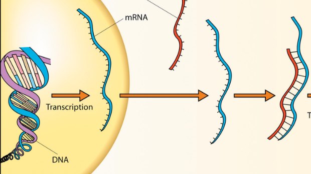
WIKIMEDIA, ROBINSON RCRISPR—a bacterial immune response best known for its genome-editing applications in the lab—has yet again been adapted for scientific purposes, this time to track RNA within cells. Researchers modified various components of CRISPR so that they could snap on to a specified messenger RNA (mRNA) within the nucleus, then enter the cytoplasm as a complex, where its fluorescent signal could be tracked.

“This work . . . sets the foundation for manipulation of the transcriptome in addition to the genome by CRISPR/Cas9,” the authors wrote in their report, published in Cell last week (March 17).

The alterations included neutering the Cas9 enzyme—which ordinarily cuts DNA—so it would grab the RNA target without cleaving it, and sticking on a fluorescent protein to allow for visual tracking. A guide RNA, along with the addition of an oligonucleotide sequence, sent the Cas9 RNA-ward.

“Just as CRISPR-Cas9 is making genetic engineering accessible to any scientist with access to basic equipment, RNA-targeted Cas9 may support countless other efforts for studying the role of RNA processing in disease or for identifying drugs that reverse defects in RNA processing,” study coauthor David Nelles of ...

Interested in reading more?

Become a Member of

The Scientist Logo
Receive full access to more than 35 years of archives, as well as TS Digest, digital editions of The Scientist, feature stories, and much more!
Already a member? Login Here

Related Topics

Meet the Author

  • kerry grens

    Kerry served as The Scientist’s news director until 2021. Before joining The Scientist in 2013, she was a stringer for Reuters Health, the senior health and science reporter at WHYY in Philadelphia, and the health and science reporter at New Hampshire Public Radio. Kerry got her start in journalism as a AAAS Mass Media fellow at KUNC in Colorado. She has a master’s in biological sciences from Stanford University and a biology degree from Loyola University Chicago.

    View Full Profile
Share
Illustration of a developing fetus surrounded by a clear fluid with a subtle yellow tinge, representing amniotic fluid.
January 2026, Issue 1

What Is the Amniotic Fluid Composed of?

The liquid world of fetal development provides a rich source of nutrition and protection tailored to meet the needs of the growing fetus.

View this Issue
Skip the Wait for Protein Stability Data with Aunty

Skip the Wait for Protein Stability Data with Aunty

Unchained Labs
Graphic of three DNA helices in various colors

An Automated DNA-to-Data Framework for Production-Scale Sequencing

illumina
Exploring Cellular Organization with Spatial Proteomics

Exploring Cellular Organization with Spatial Proteomics

Abstract illustration of spheres with multiple layers, representing endoderm, ectoderm, and mesoderm derived organoids

Organoid Origins and How to Grow Them

Thermo Fisher Logo

Products

Brandtech Logo

BRANDTECH Scientific Introduces the Transferpette® pro Micropipette: A New Twist on Comfort and Control

Biotium Logo

Biotium Launches GlycoLiner™ Cell Surface Glycoprotein Labeling Kits for Rapid and Selective Cell Surface Imaging

Colorful abstract spiral dot pattern on a black background

Thermo Scientific X and S Series General Purpose Centrifuges

Thermo Fisher Logo
Abstract background with red and blue laser lights

VANTAstar Flexible microplate reader with simplified workflows

BMG LABTECH