Culturing Drug-Resistant Tumors

Improved methods to grow patients’ tumor cells in a dish offer opportunities to find durable therapies.

Written byKerry Grens
| 1 min read

Register for free to listen to this article
Listen with Speechify
0:00
1:00
Share

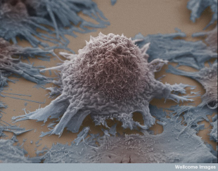
FLICKR, ANNE WESTON, LRI, CROK. WELLCOME IMAGESCancers can become resistant to drugs that once seemed like they could cure a person of the disease. In the unstopping march toward personalized medicine, researchers managed to culture drug-resistant lung tumor cells to screen them for sensitivity to various therapies, finding new and sometimes surprising drug candidates.

“It’s a substantial step,” Jeffrey Engelman of Massachusetts General Hospital Cancer Center told Time. “Because before we just had the genetic information but we wouldn’t have the cells alive so that we could test what types of therapies might work.”

Engelman and his colleagues published their study in Science last week (November 13). They succeeded in growing cells taken from 20 patients’ tumors. Nature News pointed out that, in many cases, the drug screens turned up unexpected candidates. In half of the tumors, for example, a drug that inhibits a protein called SRC managed to shrink tumor growth in vitro. “There were no genetic results that would have pointed to that combination,” Engelman told Nature News.

Engelman’s method has yet to face a clinical challenge; ...

Interested in reading more?

Become a Member of

The Scientist Logo
Receive full access to more than 35 years of archives, as well as TS Digest, digital editions of The Scientist, feature stories, and much more!
Already a member? Login Here

Related Topics

Meet the Author

  • kerry grens

    Kerry served as The Scientist’s news director until 2021. Before joining The Scientist in 2013, she was a stringer for Reuters Health, the senior health and science reporter at WHYY in Philadelphia, and the health and science reporter at New Hampshire Public Radio. Kerry got her start in journalism as a AAAS Mass Media fellow at KUNC in Colorado. She has a master’s in biological sciences from Stanford University and a biology degree from Loyola University Chicago.

    View Full Profile
Share
Illustration of a developing fetus surrounded by a clear fluid with a subtle yellow tinge, representing amniotic fluid.
January 2026, Issue 1

What Is the Amniotic Fluid Composed of?

The liquid world of fetal development provides a rich source of nutrition and protection tailored to meet the needs of the growing fetus.

View this Issue
Skip the Wait for Protein Stability Data with Aunty

Skip the Wait for Protein Stability Data with Aunty

Unchained Labs
Graphic of three DNA helices in various colors

An Automated DNA-to-Data Framework for Production-Scale Sequencing

illumina
Exploring Cellular Organization with Spatial Proteomics

Exploring Cellular Organization with Spatial Proteomics

Abstract illustration of spheres with multiple layers, representing endoderm, ectoderm, and mesoderm derived organoids

Organoid Origins and How to Grow Them

Thermo Fisher Logo

Products

nuclera logo

Nuclera eProtein Discovery System installed at leading Universities in Taiwan

Brandtech Logo

BRANDTECH Scientific Introduces the Transferpette® pro Micropipette: A New Twist on Comfort and Control

Biotium Logo

Biotium Launches GlycoLiner™ Cell Surface Glycoprotein Labeling Kits for Rapid and Selective Cell Surface Imaging

Colorful abstract spiral dot pattern on a black background

Thermo Scientific X and S Series General Purpose Centrifuges

Thermo Fisher Logo