
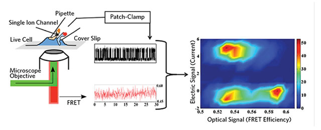
 PROBING CHANNEL CONFORMATION: Combined patch-clamp and FRET measurements reveal the configuration of ion channels. As shown in the plot of the two measured signals (above, right), channels can adopt an “open” configuration in which no ions flow (region of low FRET efficiency but also low current, bottom left). ADAPTED FROM DIBYENDU SASMAL AND H. PETER LU
As your eyes scan this page, your brain is multitasking big-time. It’s engaged in a frenetic dialog with the muscles that control your eyes; neural pathways are flickering on and off to translate words into mental pictures; and memory circuits are working to store and retrieve information.
In every case, neurons are talking to each other in the language of action potentials: rapid electrical discharges across cellular membranes that propagate from one cell to the next like electricity in a wire, driven by ion flow through neuronal membrane channels.
Traditionally, researchers have used “patch clamping” to listen in on those conversations. The technique works a bit like a cell-size voltmeter. Using a finely controlled micromanipulator, neuroscientists position an ultrathin glass pipette against the membrane of ...


