Image of the Day: Break Apart

Scientists can now view mitosis one protein (or more) at a time using a 4-D computer model.

Sukanya Charuchandra
| 1 min read

Register for free to listen to this article
Listen with Speechify
0:00
1:00
Share

The process of cell division can now be viewed with a Mitotic Cell Atlas that captures the interaction between a combination of up to seven protein types among 28 available to choose from as one cell makes two. Using this open-source, 4-D, interactive computer model, researchers can select various proteins and see how they cooperate with each other in real-time during mitosis.

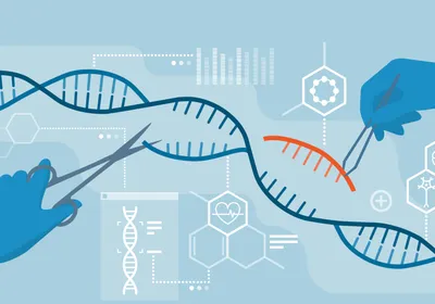
The study appeared September 10 in Nature and used CRISPR-Cas to make the proteins fluorescent in HeLa cells that were viewed under 3-D confocal microscopy. These imaging data were then coalesced into the Atlas. Several hundred more proteins that participate in mitosis need to be imaged and integrated into this system.

“Besides mitosis, the technologies developed here can be used to study proteins that drive other cellular functions, for example cell death, cell migration or metastasis of cancer cells,” coauthor Jan Ellenberg of EMBL says in a statement.“By ...

Interested in reading more?

Become a Member of

The Scientist Logo
Receive full access to more than 35 years of archives, as well as TS Digest, digital editions of The Scientist, feature stories, and much more!
Already a member? Login Here

Keywords

Meet the Author

  • Sukanya Charuchandra

    Sukanya Charuchandra

    Originally from Mumbai, Sukanya Charuchandra is a freelance science writer based out of wherever her travels take her. She holds master’s degrees in Science Journalism and Biotechnology. You can read her work at sukanyacharuchandra.com.

Share
May digest 2025 cover
May 2025, Issue 1

Study Confirms Safety of Genetically Modified T Cells

A long-term study of nearly 800 patients demonstrated a strong safety profile for T cells engineered with viral vectors.

View this Issue
iStock

TaqMan Probe & Assays: Unveil What's Possible Together

Thermo Fisher Logo
Meet Aunty and Tackle Protein Stability Questions in Research and Development

Meet Aunty and Tackle Protein Stability Questions in Research and Development

Unchained Labs
Detecting Residual Cell Line-Derived DNA with Droplet Digital PCR

Detecting Residual Cell Line-Derived DNA with Droplet Digital PCR

Bio-Rad
How technology makes PCR instruments easier to use.

Making Real-Time PCR More Straightforward

Thermo Fisher Logo

Products

fujirebio-square-logo

Fujirebio Receives Marketing Clearance for Lumipulse® G pTau 217/ β-Amyloid 1-42 Plasma Ratio In-Vitro Diagnostic Test

The Scientist Placeholder Image

Biotium Launches New Phalloidin Conjugates with Extended F-actin Staining Stability for Greater Imaging Flexibility

Leica Microsystems Logo

Latest AI software simplifies image analysis and speeds up insights for scientists

BioSkryb Genomics Logo

BioSkryb Genomics and Tecan introduce a single-cell multiomics workflow for sequencing-ready libraries in under ten hours