Image of the Day: A Swell Idea

To improve the resolution of biological samples at the cellular level, researchers inflate tissues with “swellable polymers” so that they’re easier to see under the microscope.

Written byThe Scientist
| 1 min read

Register for free to listen to this article
Listen with Speechify
0:00
1:00
Share

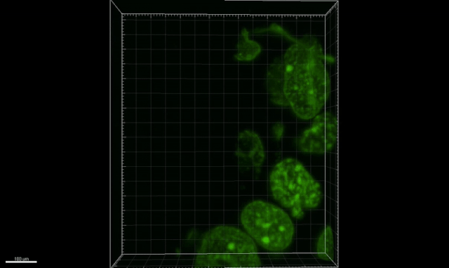
Enlarged breast cancer tissueOCTAVIAN BUCUR/LUDWIG INSTITUTE FOR CANCER RESEARCH In a study published this week (July 17) in Nature Biotechnology, Harvard and MIT scientists inflated human tissue samples of kidney minimal change disease and precancerous breast lesions to better visualize these pathologies using a technique called expansion microscopy.

"We can apply this method to any type of clinical sample and all types of human tissues, including normal and cancerous tissues," says MIT scientist and co-lead author Yongxin Zhao of MIT in a news release.

See Y. Zhao et al., “Nanoscale imaging of clinical specimens using pathology-optimized expansion microscopy,” Nature Biotechnology, doi:10.1038/nbt.3892, 2017.

Interested in reading more?

Become a Member of

The Scientist Logo
Receive full access to more than 35 years of archives, as well as TS Digest, digital editions of The Scientist, feature stories, and much more!
Already a member? Login Here
Illustration of a developing fetus surrounded by a clear fluid with a subtle yellow tinge, representing amniotic fluid.
January 2026, Issue 1

What Is the Amniotic Fluid Composed of?

The liquid world of fetal development provides a rich source of nutrition and protection tailored to meet the needs of the growing fetus.

View this Issue
Skip the Wait for Protein Stability Data with Aunty

Skip the Wait for Protein Stability Data with Aunty

Unchained Labs
Graphic of three DNA helices in various colors

An Automated DNA-to-Data Framework for Production-Scale Sequencing

illumina
Exploring Cellular Organization with Spatial Proteomics

Exploring Cellular Organization with Spatial Proteomics

Abstract illustration of spheres with multiple layers, representing endoderm, ectoderm, and mesoderm derived organoids

Organoid Origins and How to Grow Them

Thermo Fisher Logo

Products

Brandtech Logo

BRANDTECH Scientific Introduces the Transferpette® pro Micropipette: A New Twist on Comfort and Control

Biotium Logo

Biotium Launches GlycoLiner™ Cell Surface Glycoprotein Labeling Kits for Rapid and Selective Cell Surface Imaging

Colorful abstract spiral dot pattern on a black background

Thermo Scientific X and S Series General Purpose Centrifuges

Thermo Fisher Logo
Abstract background with red and blue laser lights

VANTAstar Flexible microplate reader with simplified workflows

BMG LABTECH