

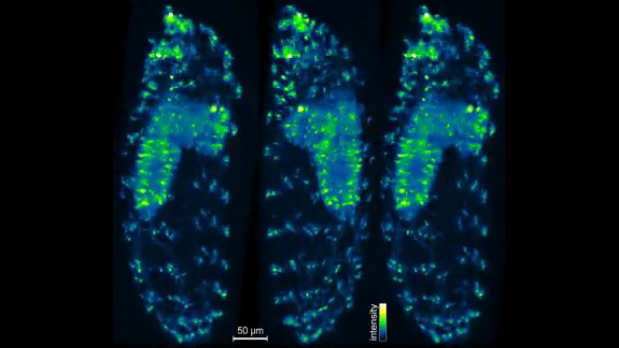
 Projections of 3-D IsoView image data of an entire Drosophila larva expressing a fluorescent indicator or neural activity through the nervous system, including the brain, ventral nerve cord, and the peripheral nervous system. (See video.)KELLER LAB, HHMI/JANELIA RESEARCH CAMPUSPhilipp Keller of the Janelia Research Campus in Ashburn, Virginia, and his colleagues today (October 26) presented a new light microscope capable of imaging large, nontransparent specimens—such as a whole fruit fly embryo—at a speed and 3-D resolution that should allow them to probe biological processes in unprecedented detail. The team’s first target: the early development of a functioning nervous system in vertebrates. The researchers described the new microscope—the IsoView, which specifically boasts improved spatial resolution over the group’s previous microscopes—in Nature Methods.
“We had decent microscopes for the type of imaging that we do—rapid imaging of cellular dynamics in large, living specimens. The temporal resolution matched the timescales of the processes we’re looking at, and we had microscopes that could give us good coverage and allow us to image for a long time without perturbing the system,” Keller said in a press release. “But we hadn’t really tried to push spatial resolution much in our microscopes to date.”
To improve resolution without compromising speed or other features of their microscopes, Keller’s group decided to collect multiple images from different angles simultaneously, using four objective lenses. While each image is still relatively low-resolution, by combining data from all the images, the researchers can reconstruct a good high-resolution view ...


