Stem Cell Alter Egos

Researchers show that cancer stem cells can exist in two distinct and interconvertible states.

Written byAbby Olena, PhD
| 2 min read

Register for free to listen to this article
Listen with Speechify
0:00
2:00
Share

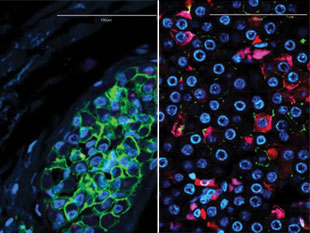
TWO-FACED: The invasive edge of a tumor (left) harbors mesenchymal-like breast cancer stem cells (green), while the interior of the tumor (right) contains epithelial-like cancer stem cells (pink).REPRINTED FROM S. LIU ET AL., STEM CELL REPORTS, 2:78-91, 2014

The paper S. Liu et al., “Breast cancer stem cells transition between epithelial and mesenchymal states reflective of their normal counterparts,” Stem Cell Reports, 2:78-91, 2014. The states Cancer metastasis is thought to rely on tumor cells losing their polarity and their connections with neighbors, enabling them to migrate and invade other tissues—an epithelial-to-mesenchymal transition. The reverse process—the mesenchymal-to-epithelial transition—then allows cells to set up shop in a new tissue. Cancer stem cells can express genes associated with both pathways, but there have been conflicting findings about the relationship between stemness and these transitions. The finding Cancer biologist Max Wicha of the University of Michigan and colleagues showed that breast cancer stem cells (BCSCs) in the same tumor can exist in both states. They found ...

Interested in reading more?

Become a Member of

The Scientist Logo
Receive full access to digital editions of The Scientist, as well as TS Digest, feature stories, more than 35 years of archives, and much more!
Already a member? Login Here

Related Topics

Meet the Author

  • abby olena

    As a freelancer for The Scientist, Abby reports on new developments in life science for the website. She has a PhD from Vanderbilt University and got her start in science journalism as the Chicago Tribune’s AAAS Mass Media Fellow in 2013. Following a stint as an intern for The Scientist, Abby was a postdoc in science communication at Duke University, where she developed and taught courses to help scientists share their research. In addition to her work as a science journalist, she leads science writing and communication workshops and co-produces a conversational podcast. She is based in Alabama.  

    View Full Profile

Published In

Share
Illustration of a developing fetus surrounded by a clear fluid with a subtle yellow tinge, representing amniotic fluid.
January 2026, Issue 1

What Is the Amniotic Fluid Composed of?

The liquid world of fetal development provides a rich source of nutrition and protection tailored to meet the needs of the growing fetus.

View this Issue
Skip the Wait for Protein Stability Data with Aunty

Skip the Wait for Protein Stability Data with Aunty

Unchained Labs
Graphic of three DNA helices in various colors

An Automated DNA-to-Data Framework for Production-Scale Sequencing

illumina
Exploring Cellular Organization with Spatial Proteomics

Exploring Cellular Organization with Spatial Proteomics

Abstract illustration of spheres with multiple layers, representing endoderm, ectoderm, and mesoderm derived organoids

Organoid Origins and How to Grow Them

Thermo Fisher Logo

Products

Brandtech Logo

BRANDTECH Scientific Introduces the Transferpette® pro Micropipette: A New Twist on Comfort and Control

Biotium Logo

Biotium Launches GlycoLiner™ Cell Surface Glycoprotein Labeling Kits for Rapid and Selective Cell Surface Imaging

Colorful abstract spiral dot pattern on a black background

Thermo Scientific X and S Series General Purpose Centrifuges

Thermo Fisher Logo
Abstract background with red and blue laser lights

VANTAstar Flexible microplate reader with simplified workflows

BMG LABTECH