Stomach Cells Change Identity to Drive Precancerous State

A study suggests that “chief” cells in the stomach act as reserve stem cells that are activated by tissue damage and may be the long-sought source of gastric cancer.

Written byDiana Kwon
| 4 min read

Register for free to listen to this article
Listen with Speechify
0:00
4:00
Share

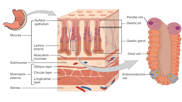
WIKIMEDIA, OPENSTAX COLLEGEIn healthy individuals, chief cells, which are found at the base of corpus glands in the stomach, act as producers of digestive enzymes. However, if the gut undergoes damage or genetic mutation, these cells have the ability to convert into stem cells that can lead to gastric cancer, according to a study published last week (June 5) in Nature Cell Biology.

“This is the first definitive demonstration that that a subset of chief cells is cancer prone and can serve as the origin of gastric cancer,” says study coauthor Nick Barker, who studies cancer and stem cells at A*STAR Institute of Medical Biology (IMB) in Singapore.

Still, the identity of gastric-cancer forming cells has been controversial, and not all researchers are convinced Barker’s study has resolved the debate.

A long-standing theory in gastroenterology is that stem cells in the stomach arise in the isthmus, the region located around the middle of the corpus glands. These cells migrate to the tops and bottoms of the glands, replenishing lost cells and maintaining homeostasis. Mutations in these stem cells, many scientists believe, can lead to precancerous lesions, such as ...

Interested in reading more?

Become a Member of

The Scientist Logo
Receive full access to more than 35 years of archives, as well as TS Digest, digital editions of The Scientist, feature stories, and much more!
Already a member? Login Here

Related Topics

Meet the Author

  • Diana is a freelance science journalist who covers the life sciences, health, and academic life. She’s a regular contributor to The Scientist and her work has appeared in several other publications, including Scientific American, Knowable, and Quanta. Diana was a former intern at The Scientist and she holds a master’s degree in neuroscience from McGill University. She’s currently based in Berlin, Germany.

    View Full Profile
Share
Illustration of a developing fetus surrounded by a clear fluid with a subtle yellow tinge, representing amniotic fluid.
January 2026

What Is the Amniotic Fluid Composed of?

The liquid world of fetal development provides a rich source of nutrition and protection tailored to meet the needs of the growing fetus.

View this Issue
Redefining Immunology Through Advanced Technologies

Redefining Immunology Through Advanced Technologies

Ensuring Regulatory Compliance in AAV Manufacturing with Analytical Ultracentrifugation

Ensuring Regulatory Compliance in AAV Manufacturing with Analytical Ultracentrifugation

Beckman Coulter Logo
Skip the Wait for Protein Stability Data with Aunty

Skip the Wait for Protein Stability Data with Aunty

Unchained Labs
Graphic of three DNA helices in various colors

An Automated DNA-to-Data Framework for Production-Scale Sequencing

illumina

Products

nuclera logo

Nuclera eProtein Discovery System installed at leading Universities in Taiwan

Brandtech Logo

BRANDTECH Scientific Introduces the Transferpette® pro Micropipette: A New Twist on Comfort and Control

Biotium Logo

Biotium Launches GlycoLiner™ Cell Surface Glycoprotein Labeling Kits for Rapid and Selective Cell Surface Imaging

Colorful abstract spiral dot pattern on a black background

Thermo Scientific X and S Series General Purpose Centrifuges

Thermo Fisher Logo