The Origins of Oversized Chromosomes

Researchers reconstruct the formation of the giant neochromosomes that contribute to some cancers.

Written byMolly Sharlach
| 2 min read

Register for free to listen to this article
Listen with Speechify
0:00
2:00
Share

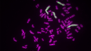
Neochromosomes (green) found in some cancer calls may be up to three times as long as normal chromosomes (magenta).MURDOCH CHILDREN’S RESEARCH INSTITUTE, OWEN MARSHALL

More than half a century ago, scientists noticed a distinctive abnormality in the karyotypes of some soft-tissue cancers: unusually large chromosomes, now referred to as neochromosomes. In a new analysis reported this week (November 10) in Cancer Cell, a team of Australian researchers have uncovered the origins of neochromosomes and revealed mechanisms that could guide therapies to block the chromosomes’ construction.

Using next-generation sequencing and mathematical modeling to investigate the development of neochromosomes, David Thomas of the Garvan Institute in Sydney and his colleagues found that the process appears to begin with the splintering and rearrangement of chromosome 12, followed by breakage-fusion-bridge cycles that lead to the amplification of oncogenes. Notably, neochromosomes often contain dozens of copies of the MDM2 and CDK4 genes, which are ...

Interested in reading more?

Become a Member of

The Scientist Logo
Receive full access to more than 35 years of archives, as well as TS Digest, digital editions of The Scientist, feature stories, and much more!
Already a member? Login Here

Related Topics

Meet the Author

Share
Illustration of a developing fetus surrounded by a clear fluid with a subtle yellow tinge, representing amniotic fluid.
January 2026, Issue 1

What Is the Amniotic Fluid Composed of?

The liquid world of fetal development provides a rich source of nutrition and protection tailored to meet the needs of the growing fetus.

View this Issue
Skip the Wait for Protein Stability Data with Aunty

Skip the Wait for Protein Stability Data with Aunty

Unchained Labs
Graphic of three DNA helices in various colors

An Automated DNA-to-Data Framework for Production-Scale Sequencing

illumina
Exploring Cellular Organization with Spatial Proteomics

Exploring Cellular Organization with Spatial Proteomics

Abstract illustration of spheres with multiple layers, representing endoderm, ectoderm, and mesoderm derived organoids

Organoid Origins and How to Grow Them

Thermo Fisher Logo

Products

Brandtech Logo

BRANDTECH Scientific Introduces the Transferpette® pro Micropipette: A New Twist on Comfort and Control

Biotium Logo

Biotium Launches GlycoLiner™ Cell Surface Glycoprotein Labeling Kits for Rapid and Selective Cell Surface Imaging

Colorful abstract spiral dot pattern on a black background

Thermo Scientific X and S Series General Purpose Centrifuges

Thermo Fisher Logo
Abstract background with red and blue laser lights

VANTAstar Flexible microplate reader with simplified workflows

BMG LABTECH