A Twist of Fate

Once believed to be irrevocably differentiated, mature cells are now proving to be flexible, able to switch identities with relatively simple manipulation.

Written byJonathan Slack
| 12 min read

Register for free to listen to this article
Listen with Speechify
0:00
12:00

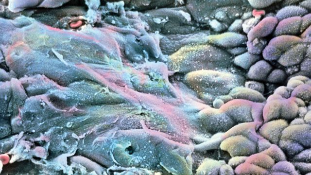
THE LONG AND SHORT OF IT: The epithelium of the cervix consists of both flattened squamous cells (left, top), which occur in many layers and slough off as they are replaced by cells beneath them, and columnar cells (left, bottom), which form a mucus-secreting monolayer covered in microvilli.© P.M. MOTTA & S. MAKABE/SCIENCE SOURCE IMAGES

In 1998, University of Bath biologist David Tosh had a eureka moment. He noticed that some of the rat pancreatic exocrine cells he was working with had become unusually large and flat. After further testing, he identified the cause of the abnormality: the cells, which belonged to an established line called AR42j, were no longer pancreatic cells at all; they were hepatocytes, the principal cell type of the liver. The cells had changed their identity simply under the influence of the synthetic hormone dexamethasone, which had been added to the medium to enhance endocrine cell secretions.1

This wasn’t the first demonstration that cells could change type. Pathologists had long reported the transformation of one cell type into another in humans, a normally harmless process known ...

Interested in reading more?

Become a Member of

The Scientist Logo
Receive full access to digital editions of The Scientist, as well as TS Digest, feature stories, more than 35 years of archives, and much more!
Already a member?
Add The Scientist as a preferred source on Google

Add The Scientist as a preferred Google source to see more of our trusted coverage.

Meet the Author

Related Topics

Published In

Related articles background image
The Scientist Digest cover September 2026
September 2026

Multiplex Microscopy Becomes Easier with Encoded Antibodies

A new system that enables researchers to uniquely tag monoclonal antibodies for use in microscopy could help simplify complex imaging studies.

View this Issue
Essential Genes Are Dominantly Activated by Single Transcription Factors

Essential Genes Are Dominantly Activated by Single Transcription Factors

EpiCypher Logo
Rethinking ALS Biomarkers: From Discovery to Clinical Impact

Rethinking ALS Biomarkers: From Discovery to Clinical Impact

Alamar Biosciences logo
Engineering CAR-Neutrophils In Vivo to Target Glioblastoma

Engineering CAR-Neutrophils In Vivo to Target Glioblastoma

Miltenyi
Best Practices for qPCR Assay Design and Optimization

Best Practices for qPCR Assay Design and Optimization

Bio-Rad

Products

Closeup image of a multi channel pipette dispensing pink liquid into a 96-well plate.

The ASSIST PLUS pipetting robot for affordable workflow automation

Integra Logo
Single cells in suspension

Rapidly isolate primary cells and make uniform single-cell suspensions with Corning® Cell Strainers

Corning logo
Abstract image representing cell membranes linked together.

CellBrite® Steady Membrane Stain: Cell surface staining built for real-time imaging

Biotium
sino biological logo

Monod Bio Licenses AI-designed Protein Technologies to SignalChem Biotech for Custom Discovery Assays