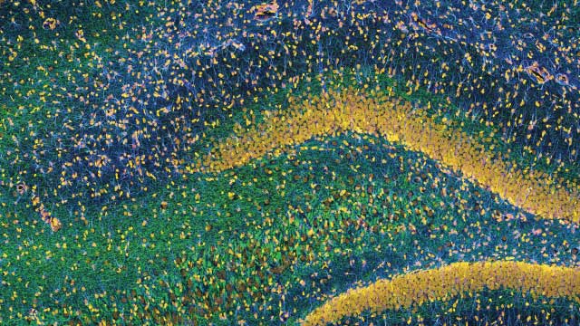
IT TAKES A VILLAGE: Glia (cyan) in the central nervous system are normally considered support for neurons, but research is revealing how these cells can contribute to the aberrant firing of pain pathways. (Rat hippocampus shown here. Neurofilaments in green; DNA in yellow.)TOM DEERINCK, NATIONAL CENTER FOR MICROSCOPY AND IMAGING RESEARCH

When someone is asked to think about pain, he or she will typically envision a graphic wound or a limb bent at an unnatural angle. However, chronic pain, more technically known as persistent pain, is a different beast altogether. In fact, some would say that the only thing that acute and persistent pain have in common is the word “pain.” The biological mechanisms that create and sustain the two conditions are very different.

Pain is typically thought of as the behavioral and emotional results of the transmission of a neuronal signal, and indeed, acute pain, or nociception,...

Long considered to be little more than cellular glue holding the brain together, glia, which outnumber neurons 10 to 1, are now appreciated as critical contributors to the health of the central nervous system, with recognized roles in the formation of synapses, neuronal plasticity, and protection against neurodegeneration. And over the past 15 to 20 years, pain researchers have also begun to appreciate the importance of these cells. Research has demonstrated that glia seem to respond and adapt to the cumulative danger signals that can result from disparate kinds of injury and illness, and that they appear to prime neural pathways for the overactivation that causes persistent pain. In fact, glial biology may hold important clues to some of the mysteries that have perplexed the pain research field, such as why the prevalence of persistent pain differs between the sexes and why some analgesic medications fail to work.

Importantly, these insights are not just going from the bench to the bookshelf. Rather, large pharmaceutical companies have taken an interest in translating new glia-targeting therapies to the clinic to treat persistent pain, a malady that costs society more than cancer, heart disease, and diabetes combined. A wealth of preclinical evidence supports this translational potential. Every relevant animal and cell model of persistent pain tested to date shows histological and molecular signs of changed glial activity or pharmacological sensitivity to drugs that target these cells. With several new chemical entities and drugs being repurposed to target glia for pain, and with continued laboratory work interrogating the mechanisms underlying glia’s involvement in persistent pain processing, the field is poised to make strides in both understanding and treating this enigmatic ailment.

TWO PAIN PATHS DIVERGE IN THE BODY: The acute pain that results from injury or disease, also known as nociception, is very different from the persistent pain that continues even after the damage is resolved, or in some cases, in the absence of an acute injury in the first place. The difference, researchers are learning, comes down to the neural mechanisms that trigger these distinct signals in the brain.
See full infographic: WEB | PDF
© THOM GRAVES. REDRAWN FROM FIGURE 1 IN BIOL PSYCHIATRY, 65:732-741, 2009.

The neuroimmune brain

Until very recently, the brain and spinal cord were thought to be shielded from the body’s immune system. But evidence to the contrary has been accumulating for years. And recently, researchers discovered the central nervous system’s lymphatic system, which traffics thousands of peripheral immune cells into and out of the healthy brain.2 This recognition of the immunocompetence of the central nervous system validates a long-appreciated idea—that in some persistent pain states, peripheral immune cells contribute to the function of somatosensory synapses in the brain and spinal cord. In addition, although they are not themselves considered classical immune cells, glia—which comprise a range of phenotypically different cell types, including astrocytes, microglia, and oligodendrocytes—perform a role similar to that of the peripheral immune system, and can also contribute to exaggerated pain responses.

See “Immune System Maintains Brain Health

While synapses were once thought to involve just two participants—the pre- and postsynaptic neuronal terminals—researchers now recognize that upward of 90 percent of neural connections include one, two, and sometimes even three additional types of cellular players.

Glial biology may hold important clues to some of the mysteries that have perplexed the pain research field.

In persistent pain, if glial function is modified in and around synapses, the transmission of nociceptive signals can be augmented in a way that will result in exaggerated pain responses. For example, projections from astrocytes known as endfeet closely monitor synaptic activity for changes in neuronal firing. When the glial cells detect an increase in the extracellular concentrations of neurotransmitters, they begin to take up greater amounts of the molecules in an attempt to bring the hyperactive synapses under control. Under states of persistent pain, however, there is a significant downregulation of the molecular transporters on astrocytes that are responsible for maintaining excitatory neurotransmitter homeostasis, resulting in less removal of excess excitatory neurotransmitters.

Microglia, meanwhile, survey the synaptic space for local and distant paracrine signals such as cytokines, chemokines, and trophic factors that drive neuronal adaptations at the level of the synapse to continue to refine their likelihood of firing.3 Some glial cells also release their own proinflammatory cytokines and other mediators, such as reactive oxygen and nitrogen species. Along with additional proinflammatory factors from peripheral immune cells, these compounds can prime the synapse for heightened neuronal firing by increasing the release of excitatory neurotransmitters from neurons.

In addition, glial cytokines and chemokines are known to drive increased production of neuronal receptors that the neurotransmitters bind to on the postsynaptic terminal, as well as the modification of receptor subunits, to promote a state of enhanced neuroexcitability and, therefore, pain sensitivity. And if all that weren’t bad enough, these glial interactions also cause a loss of inhibitory control measures in somatosensory neuronal networks, further heightening and spreading nociceptive signal transmission.

Astrocytes, such as these human cells growing in culture, are but one of an array of the glia, which greatly outnumber neurons.COURTESY OF CNBP RESEARCH FELLOW LINDSAY PARKER, MACQUARIE UNIVERSITY

It is abundantly clear that glia can enhance the firing of neurons in pain-sensing pathways to promote exaggerated responses. But how important to persistent pain are misbehaving glia? In 2016, Linda Watkins and Peter Grace of the University of Colorado Boulder and their colleagues answered this question, using a new technology known as designer receptors exclusively activated by designer drugs (DREADDs). Watkins, Grace, and their colleagues constructed an exclusively microglia-targeting viral vector that would introduce into rats an engineered mutant form of a G protein–coupled receptor that can only be activated by the DREADD-selective ligand clozapine-N-oxide (CNO). Injecting CNO, the researchers observed the activation of microglial proinflammatory responses and surmised that this response was sufficient to elicit heightened pain in the animals, even in the absence of neuronal injury.4

Hence, glia are critical to the exaggeration of pain signals that results from aberrant neuronal firing—but these immune-like cells appear capable of triggering persistent pain symptoms on their own, at least in animal models. And recent neuroimaging studies by Harvard University’s Marco Loggia and colleagues provide the first evidence that the extent of glial reactivity may be related to the severity of persistent pain in humans. The researchers employed integrated positron emission tomography–magnetic resonance imaging and a recently developed radioligand that binds to the glial translocator protein (TSPO), an anti-inflammatory molecule whose upregulation is thought to be triggered by periods of heightened glial activity to control local inflammation and reduce pain. Indeed, the team found in patients with chronic lower back pain that increased TSPO levels in the thalamus, a key higher brain region in the somatosensory pathway, negatively correlated with clinical pain scores as well as with circulating levels of the proinflammatory cytokine interleukin-6.5 These data indirectly support the role of glia signaling in persistent pain, not just in animal models but in humans as well, and provide hope that if we can find a way to regain control over glial hyper-responsiveness, we may be able to develop an effective treatment.

Glial mediators

Now that we know glia can modulate aberrant pain responses caused by somatosensory dysfunction, and can even misbehave on their own to drive persistent pain, we can ask what signals these cells are responding to. While some such signals are well known, mechanisms governing glial involvement in pain processing remain to be discovered.

In early 2016, Allan Basbaum of the University of California, San Francisco School of Medicine led a team that identified a fascinating mechanism by which sensory neurons communicate danger to the spinal cord. The team discovered that colony-stimulating factor 1 (CSF1) is produced inside mouse primary sensory neurons after injury and that the protein is physically transported to the spinal cord along the neuronal axon. Once at the heart of the spinal somatosensory processes, this neuronal payload is released to selectively target the microglial CSF1 receptor (CSF1R), triggering a cascade of signaling events that drive microglial cell proliferation and enhanced inflammatory cytokine production, and, as a result, an exaggerated pain response in the animals.6

Pharmaceutical com­panies have taken an interest in translat­ing new glia-targeting therapies to the clinic to treat persistent pain.

Another mediator of glial contributions to persistent pain appears to be the pattern-recognition receptor systems of the innate immune system, which detect conserved features in and on invading pathogens. The Toll-like receptor (TLR) system can trigger an immune response after detecting a wide range of molecular patterns—called pathogen-associated molecular patterns (PAMPs)—on invading viral, fungal, and bacterial species. This same receptor system also enables communication with the microbiome, via detection of so-called microbiome-associated molecular patterns (MAMPs). Under persistent pain conditions, however, this detection system is upregulated on glia in the somatosensory system, and is activated by endogenous signals from stressed or damaged cells termed danger-associated molecular patterns (DAMPs). The consequences of DAMP-induced microglial TLR activation serve as a first-line trigger of microglial inflammatory cytokines, which can initiate a cascade that drives heightened pain responses.

Based on the role that glia play in exaggerated pain states, the last thing that a doctor would want to do to a persistent pain patient is heighten the reactivity of the glial innate immune pattern recognition systems, or increase proinflammatory signals. But evidence has accrued over the last two decades that opioids—the gold standard for the management of acute and cancer pain, and increasingly used for the management of chronic pain—drive precisely this unwanted reactive phenotype in spinal glia and cause increased and protracted pain sensitivity. And recent research points to the innate immune system’s involvement in the unwanted side effects of opioid analgesics, with preclinical and clinical studies implicating glial responses in both tolerance and dependence. Additionally, the presentation of opioid-induced hyperalgesia—heightened pain following exposure to opioids—has been linked to glial responses triggered by the activation of pattern recognition receptors by what’s termed xenobiotic-associated molecular patterns (XAMPs). These new discoveries will soon force us to change the way we use opioids and to consider new approaches to treating persistent pain.

Astrocytes (brown mat below a cultured network of neurons shown in tan) can remove neurotransmitters as they cross synapses (round tan balls in top image), affecting the likelihood that postsynaptic neurons will fire. Under states of persistent pain, however, astrocytes are less effective at taking up excess neurotransmitters.NATIONAL CENTER FOR MICROSCOPY AND IMAGING RESEARCHAstrocytes in cultureCOURTESY OF CNBP RESEARCH FELLOW PHLLIPP REINECK, RMIT; HAND: © ISTOCK.COM/HORILLAZ

Glial-targeted therapies

The growing recognition that glia are key players in persistent pain has raised interest in mining this system for novel targets to alleviate the condition. So far, however, glial-targeted treatments have fallen short. In fact, the 2009 failure of a closely watched Phase 2 clinical trial of propentofylline—a glial modulator that had shown efficacy for treating persistent pain in animal models—all but killed off drug development in this space for years.

The lack of a successful glial-targeted therapy thus far can be attributed to a couple of key factors. First is the lack of a translational rodent-to-human experimental model that captures the mechanisms underpinning persistent pain, including both neuronal and glial components. Indeed, ongoing exploration into the 2009 trial, which failed to reduce pain in patients with post-herpetic neuralgia, a complication of shingles, pointed to differences in how human and rodent microglia responded to propentofylline in vitro.7 Secondly, the field still lacks objective biomarkers of persistent pain that allow for enriched subject recruitment into trials and objective quantification of the pain experience.

Sex differences in pain processing also complicate the search for effective glial-targeted analgesics. While healthy males and females don’t have substantial differences in sensitivity to acute pain, women are significantly more susceptible than men to persistent pain. The field has yet to reach a consensus on the underlying reason for this observation, but one possible culprit is variation between the sexes in the activity of the immune cell types responsible for the creation and maintenance of persistent pain. Scientists will need to be conscious of this difference and, when it comes to the application of glial-targeted therapies, consider developing sex-specific treatment approaches for persistent pain in men and women.

See “Sex Differences in Opioid Analgesia: A Complicated Picture?

More broadly, the new glial view of persistent pain is also changing the way we think about other prevalent pain conditions. Classically viewed peripheral inflammatory pathologies associated with persistent pain, such as rheumatoid arthritis and osteoarthritis, are now being recognized as having previously unexplored central nervous system glial contributions. In these conditions, disease-modifying antirheumatic drugs are successfully blocking the peripheral manifestation of the debilitating diseases. However, clinical data demonstrate that exaggerated and persistent pain continues in the absence of any ongoing disease progression, and recent preclinical rodent studies have demonstrated that this persistent pain has its origins in the central nervous system and can be controlled by glial-targeted therapies. For example, spinal delivery of glial-targeted drugs diminished pain behaviors in rodent models of rheumatoid arthritis and osteoarthritis.8

No one can question that persistent pain is a very complex, multicellular disease state. There is no neuronal loss or profound lesion; the triggering injury or disease has resolved, or never existed peripherally in the first place. As a result, often nothing can be measured histologically or via blood tests, leading some medical professionals to incorrectly tell their patients that “the pain is all in your head.” But the lack of a definitive clinical test for persistent pain does not mean that the condition has no biological basis. Indeed, we now know that an alteration in the homeostasis of multiple cellular systems changes the reactivity of somatosensory pain pathways, causing persistent, unremitting agony. Pain physician and pharmacologist Paul Rolan of the University of Adelaide calls this condition a “cancer of the soul.”

But there is hope. Despite the clinical failures to date, recent breakthroughs point toward new molecular players in the glial-driven persistence of pain, and researchers continue to pursue drugs that target these mechanisms. Along with continued research into the biological basis of persistent pain, these efforts may one day lead to disease-modifying treatments for a painful and costly medical problem. 

Mark R. Hutchinson is the director of the Australian Research Council Centre of Excellence for Nanoscale BioPhotonics at the University of Adelaide Medical School.

References

  1. M.M. Buchanan et al., “Toll–like receptor 4 in CNS pathologies,” J Neurochem, 114:13-27, 2010.
  2. A. Louveau et al., “Structural and functional features of central nervous system lymphatic vessels,” Nature, 523:337-41, 2015.
  3. R.-R. Ji et al., “Pain regulation by non-neuronal cells and inflammation,” Science, 354:572-77, 2016.
  4. P. Grace et al., “Selective manipulation of spinal microglia by chemogenetics: Implications for allodynia and inflammatory signaling,” J Pain, 16:S38, 2015.
  5. M.L. Loggia et al., “Evidence for brain glial activation in chronic pain patients,” Brain, 138:604-15, 2015.
  6. Z. Guan et al., “Injured sensory neuron–derived CSF1 induces microglial proliferation and DAP12-dependent pain,” Nat Neurosci, 19:94-101, 2016.
  7. R.P. Landry et al., “Propentofylline, a CNS glial modulator does not decrease pain in post-herpetic neuralgia patients: In vitro evidence for differential responses in human and rodent microglia and macrophages,” Exp Neurol, 234:340-50, 2012.
  8. C.A. Christianson et al., “Characterization of the acute and persistent pain state present in K/BxN serum transfer arthritis,” Pain, 151:394-403, 2010.

Interested in reading more?

Magaizne Cover

Become a Member of

Receive full access to digital editions of The Scientist, as well as TS Digest, feature stories, more than 35 years of archives, and much more!