
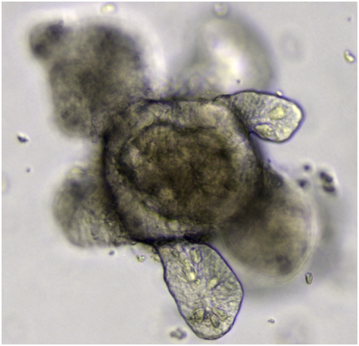
 Intestinal organoidWIKIMEDIA, MERITXELL HUCHTraditionally, gene therapy efforts have attempted to treat genetic diseases by modifying DNA inside a patient’s body, but it has been a challenge to deliver the genetic material to all the target tissue, let alone to do so safely. But in recent years, advances in gene editing and stem cell research have enabled scientists to correct genetic defects in a patient’s own cells and grow tissue-specific “organoids” in vitro. These mini organs hold promise for modeling disease, screening drugs, and—potentially—replacing defective tissue in patients.
Advances in CRISPR/Cas9 gene-editing have enabled researchers to easily and accurately make genetic modifications to human DNA. Meanwhile, the ability to reprogram cells into induced pluripotent stem cells (iPSCs) and other advances in tissue engineering have enabled scientists to grow a range of different tissues, including mini guts, kidneys, and brains.
“Both the CRISPR technology and organoid technology are relatively recent developments,” Benjamin Freedman of the University of Washington told The Scientist. “Genetics is making it possible now to understand—on an individual basis—where a disease is coming from. Combine that with the ability to go to a specific place that’s causing disease and correct it and put that tissue back into a patient, and you have a really powerful combination of tools.”
Hans Clevers, a ...


