Unmasking Secret Identities

A tour of techniques for measuring DNA hydroxymethylation

Written byKate Yandell
| 9 min read

Register for free to listen to this article
Listen with Speechify
0:00
9:00
Share

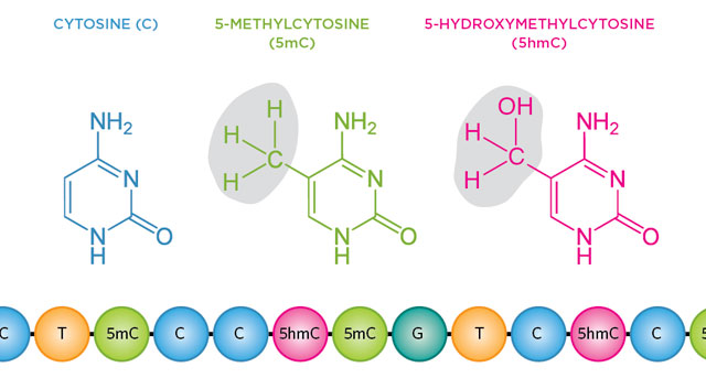
The DNA base cytosine has a tendency to play dress-up, gaining and shedding chemical modifications. For more than 40 years, scientists have known that methyl groups attached to cytosine’s fifth carbon atom can alter gene expression. These epigenetically marked bases, called 5-methylcytosines (5mCs), help to determine how hundreds of cell types in the human body differentiate and maintain their identities, despite having the same genetic backgrounds.

Recently, researchers have rediscovered a mostly ignored epigenetic variant that results when a methyl group on a cytosine takes on a hydroxyl group to form 5-hydroxymethylcytosine (5hmC). The favored method for detecting methylation is bisulfite sequencing, which converts unmodified cytosine to uracil, which then reads as thymine following PCR amplification. Modified cytosines continue to read as cytosines. This technique ...

Interested in reading more?

Become a Member of

The Scientist Logo
Receive full access to digital editions of The Scientist, as well as TS Digest, feature stories, more than 35 years of archives, and much more!
Already a member? Login Here
Illustration of a developing fetus surrounded by a clear fluid with a subtle yellow tinge, representing amniotic fluid.
January 2026, Issue 1

What Is the Amniotic Fluid Composed of?

The liquid world of fetal development provides a rich source of nutrition and protection tailored to meet the needs of the growing fetus.

View this Issue
Skip the Wait for Protein Stability Data with Aunty

Skip the Wait for Protein Stability Data with Aunty

Unchained Labs
Graphic of three DNA helices in various colors

An Automated DNA-to-Data Framework for Production-Scale Sequencing

illumina
Exploring Cellular Organization with Spatial Proteomics

Exploring Cellular Organization with Spatial Proteomics

Abstract illustration of spheres with multiple layers, representing endoderm, ectoderm, and mesoderm derived organoids

Organoid Origins and How to Grow Them

Thermo Fisher Logo

Products

Brandtech Logo

BRANDTECH Scientific Introduces the Transferpette® pro Micropipette: A New Twist on Comfort and Control

Biotium Logo

Biotium Launches GlycoLiner™ Cell Surface Glycoprotein Labeling Kits for Rapid and Selective Cell Surface Imaging

Colorful abstract spiral dot pattern on a black background

Thermo Scientific X and S Series General Purpose Centrifuges

Thermo Fisher Logo
Abstract background with red and blue laser lights

VANTAstar Flexible microplate reader with simplified workflows

BMG LABTECH