Explaining Elephants’ Cancer Resistance

Two studies reveal that the giant mammals have dozens of extra copies of a cancer-preventing gene.

Written byJef Akst
| 2 min read

Register for free to listen to this article
Listen with Speechify
0:00
2:00
Share

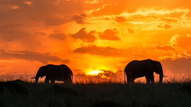
WIKIMEDIA, CHRISTOPHER MICHELWith about 100 times as many cells as a human, a 4,800-kilogram African elephant should, in theory, be more prone to accruing mutations that cause one of those cells to grow out of control. But elephants and other large mammals have surprisingly low rates of cancer, despite living for decades. As Joshua Schiffman, a pediatric oncologist at the University of Utah’s Huntsman Cancer Institute in Salt Lake City, and his colleagues found in a paper published last week (October 8) in JAMA, fewer than 5 percent of captive elephants worldwide die from cancer.

“Long-lived animals with lots of cells should all be dropping dead of cancer,” Schiffman told Science. “But they don’t or they’d go extinct.”

To understand why elephants are apparently cancer resistant, Schiffman and his colleagues scoured the genome of an African elephant and found 40 copies of the p53 gene, known to be important in cancer prevention. The Asian elephant genome contains between 30 and 40 copies of p53. Humans harbor only two copies of the gene. But elephant cells were no better at repairing DNA damage than human cells, in vitro experiments revealed. Instead, the researchers suggested that p53 helps elephants kill off precancerous cells before they become problematic.

Another study published last week (October 6) on the preprint server BioRxiv also found dozens ...

Interested in reading more?

Become a Member of

The Scientist Logo
Receive full access to more than 35 years of archives, as well as TS Digest, digital editions of The Scientist, feature stories, and much more!
Already a member? Login Here

Related Topics

Meet the Author

  • Jef (an unusual nickname for Jennifer) got her master’s degree from Indiana University in April 2009 studying the mating behavior of seahorses. After four years of diving off the Gulf Coast of Tampa and performing behavioral experiments at the Tennessee Aquarium in Chattanooga, she left research to pursue a career in science writing. As The Scientist's managing editor, Jef edited features and oversaw the production of the TS Digest and quarterly print magazine. In 2022, her feature on uterus transplantation earned first place in the trade category of the Awards for Excellence in Health Care Journalism. She is a member of the National Association of Science Writers.

    View Full Profile
Share
Illustration of a developing fetus surrounded by a clear fluid with a subtle yellow tinge, representing amniotic fluid.
January 2026, Issue 1

What Is the Amniotic Fluid Composed of?

The liquid world of fetal development provides a rich source of nutrition and protection tailored to meet the needs of the growing fetus.

View this Issue
Skip the Wait for Protein Stability Data with Aunty

Skip the Wait for Protein Stability Data with Aunty

Unchained Labs
Graphic of three DNA helices in various colors

An Automated DNA-to-Data Framework for Production-Scale Sequencing

illumina
Exploring Cellular Organization with Spatial Proteomics

Exploring Cellular Organization with Spatial Proteomics

Abstract illustration of spheres with multiple layers, representing endoderm, ectoderm, and mesoderm derived organoids

Organoid Origins and How to Grow Them

Thermo Fisher Logo

Products

Brandtech Logo

BRANDTECH Scientific Introduces the Transferpette® pro Micropipette: A New Twist on Comfort and Control

Biotium Logo

Biotium Launches GlycoLiner™ Cell Surface Glycoprotein Labeling Kits for Rapid and Selective Cell Surface Imaging

Colorful abstract spiral dot pattern on a black background

Thermo Scientific X and S Series General Purpose Centrifuges

Thermo Fisher Logo
Abstract background with red and blue laser lights

VANTAstar Flexible microplate reader with simplified workflows

BMG LABTECH